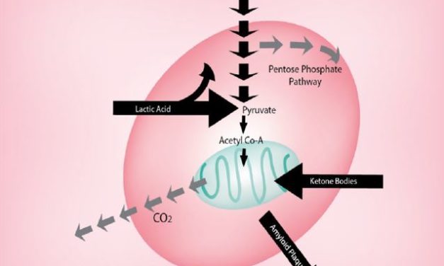
Résistance à la circulation des savoirs scientifiques : l’effet Warburg en cancérologie.

Résistance à la circulation des savoirs scientifiques : l’effet Warburg en cancérologie.

Novembre 2018
Conférence: 14èmes Journées du Cancéropôle Grand Sud-Ouest
Projet: Historical analysis & scientometric modeling of sleeping beauty and serendipity discoveries.

Auteurs:

Philippe Gorry           University of Bordeaux
Pascal Ragouet           University of Bordeaux
andrew lumsden

Résumé :

À l’heure actuelle, les notions d’innovation et de recherche translationnelle sont très fréquemment utilisées. Elles renvoient souvent à la nécessité de contribuer à une circulation de connaissances scientifiques nouvelles dans une logique de valorisation. Pourtant, la nouveauté n’est pas exclusivement liée à des découvertes : des travaux peuvent rester très longtemps ignorés, avant d’être reconnus.
Ce phénomène de reconnaissance tardive connaît un regain d’intérêt avec l’analyse en scientométrie des « belles au bois dormant » (ou « sleeping beauties »), et de leur prévalence et importance en histoire et sociologie des sciences (Gorry & Ragouet, 2016 ; El Aichouchi & Gorry, 2018).
Cette notion introduite par Van Raan (2004) désigne un article très peu cité pendant une période initiale de plus de 10 ans (période du « sommeil »), qui reçoit au cours des années suivantes beaucoup de citations (période du « réveil ») du fait de son utilisation dans la publication d’un chercheur (le « baiser du Prince »). Le travail présenté se propose d’explorer les dimensions scientométriques, historiques et sociologiques du sommeil et du réveil des travaux d’Otto Warburg. Ce chercheur allemand du début du XXe siècle a été récompensé du prix Nobel en 1931 « pour sa découverte de la nature et du mode d’action de l’enzyme respiratoire », dans les plantes et les cellules cancéreuses. Il postulera l’hypothèse que les cellules tumorales n’ont pas besoin d’un milieu riche en oxygène pour se développer.
Fort de ses résultats, il ira même plus loin dans ses conclusions, et recommandera un régime nutritionnel pour prévenir et guérir du cancer. Ces propos déclencheront alors une forte polémique. Le métabolisme des cellules cancéreuses décrit par Warburg est devenu depuis un éponyme associé au cancer : l’« effet Warburg », et constitue depuis ces dernières années une nouvelle piste thérapeutique.
Sur le plan bibliométrique, l’article fondateur de Warburg à propos du lien entre métabolisme et cancer, publié en 1956 dans la prestigieuse revue Science, remplit les critères de Van Raan. Cet article a traversé un sommeil de près de 45 ans avec moins de 10 citations annuelles avant d’être réveillé à partir de l’année 2006 pour atteindre rapidement en 2016 un pic maximum de 686 citations, marquant la reconnaissance étendue des travaux de Warburg. Afin de comprendre la dynamique du réveil, nous avons eu recours à une analyse du réseau des citations de ce travail afin d’identifier les « Princes », ces chercheurs ayant contribué significativement à la reconnaissance de l’importance des travaux de Warburg.
Cela nous a permis ainsi d’identifier des chercheurs en activité ayant participé à ce réveil, ce afin de mener une série d’entretiens semi-directifs portant, d’une part, sur leur trajectoire et, d’autre part, sur le contexte dans lequel ils ont été amenés à citer le travail d’Otto Warburg, les raisons qui les ont poussés à redécouvrir sa contribution.
Les entretiens portent notamment sur la façon dont les chercheurs qui citent le travail de Warburg plus ou moins longtemps après son réveil se représentent cette contribution, sur ce qu’ils considèrent comme ses apports, sur l’importance de ce que l’on appelle maintenant l’« effet Warburg », sur le caractère éventuellement controversé de la thèse et, le cas échéant, sur les échanges que ces chercheurs ont eus avec des cliniciens à propos du travail de Warburg. Le travail proposé ici et qui repose sur des méthodes mixtes vise par conséquent à explorer, au sein de la recherche française, les facteurs explicatifs de la reconnaissance tardive du travail de Warburg et d’amorcer, à partir de cette étude de cas, une exploration des mécanismes qui pourrait expliquer la résistance à la circulation de résultats et savoirs scientifiques et à leur transformation en pratiques diagnostiques et thérapeutiques dans le domaine de la cancérologie.


L’exportation d’entropie comme force motrice de l’évolution

L’exportation d’entropie comme force motrice de l’évolution


Citation: M. Henry, L. Schwartz (2019) Entropy export as the driving force of evolution. Substantia 3(2) Suppl. 3: 29-56. doi: 10.13128/Substantia-324

Copyright: © 2019 JM. Henry, L.Schwartz . l s’agit d’un article en accès libre, évalué par les pairs, publié par Firenze University Press (http://www.fupress.com/substantia) et distribué selon les termes de la licence Creative Commons Attribution, qui permet une utilisation, une distribution et une reproduction sans restriction sur n’importe quel support, à condition que l’auteur original et la source soient crédités.

Déclaration de disponibilité des
données : Toutes les données pertinentes se trouvent dans l’article et dans les fichiers d’informations complémentaires.

Intérêts concurrents : Le(s) auteur(s) ne déclare(nt) aucun conflit d’intérêt.

Source (en anglais):
https://riviste.fupress.net/index.php/subs/article/view/324/312

Marc Henry, Laurent Schwartz*

Assistance Publique des Hôpitaux de Paris, Paris, France
*Auteur correspondant : dr.laurentschwartz@gmail.com

Résumé. Le concept d’entropie a été forgé au milieu du XIXe siècle pour prédire comment un système chimique peut subir une évolution spontanée dans le temps. A l’aube du vingtième siècle, quatre principes d’une nouvelle science appelée « thermodynamique » ont été fermement établis. Il s’agit du concept d’équilibre thermique (loi zéro), de la conservation de l’énergie (première loi), de l’augmentation spontanée de l’entropie au cours du temps (deuxième loi) et de la disparition de l’entropie au zéro absolu de température (troisième loi également appelée théorème de Nernst). Parmi ces principes, le plus gênant est la deuxième loi qui désigne les gaz comme le produit final de toute évolution dans un système fermé. D’après l’observation directe, il semble que les systèmes biologiques subissent une évolution spontanée, passant de gaz caractérisés par une entropie maximale à des structures hautement complexes affichant des entropies considérablement plus faibles, ce qui constitue une violation apparente de la deuxième loi. Cependant, il est aussi parfaitement permis de considérer la Terre comme un système ouvert capable de subir une diminution spontanée (locale) de l’entropie, à condition que l’entropie excédentaire puisse être exportée efficacement vers l’univers entier par le biais de photons infrarouges invisibles. À condition que l’entropie exportée sous forme de rayonnement infrarouge invisible soit beaucoup plus élevée que la diminution d’entropie observée sur la Terre, l’apparition de la vie sur cette planète devient pleinement conforme aux lois de la thermodynamique. Les conséquences d’un tel point de vue élargi prenant en compte tous les types de processus (réversibles et irréversibles) sont étudiées en profondeur dans cet article. Nous soutenons ici que la première condition permettant l’apparition spontanée de la vie sur Terre est l’existence d’un métabolisme, prenant la forme de cycles thermodynamiques capables de générer une grande quantité d’entropie en dégradant des systèmes moléculaires à faible entropie (nourriture) en composés moléculaires à haute entropie (déchets). Deux cycles possibles ont été identifiés, s’appuyant sur la très faible entropie du noyau métallique de la Terre pour générer des gaz réducteurs (tels que H2, CO, NH3, H2S) couplés à la faible entropie du rayonnement solaire (nourriture) et produisant des minéraux (tels que le serpentin, le sulfure métallique, la magnétite, la goethite) ainsi que des gaz à haute entropie (eau, dioxyde de carbone) comme déchets. L’important flux d’entropie généré par ces processus peut alors être utilisé pour construire des systèmes moléculaires à faible entropie basés sur des espèces carbonées réduites et des phosphates solubles que l’on observe dans toute cellule vivante. Une autre conséquence de cette approche est de souligner le rôle omniprésent joué par l’eau dans chaque caractéristique de la vie, à travers le concept d’ « activité de l’eau ».

lire la suite …


Manifesto for an Effective Treatment of the Diseases

Manifesto for an Effective Treatment of the Diseases

Contact : dr.laurentschwartz@gmail.com


To all those who had the courage to choose and therefore to pay the price

Warning

You think you know cancer. You are hoping your doctor is getting this disease under control. It is not so. Cancer is still largely a “terra incognita”. This text is the result of long reflections and experiments carried out by many physicians, researchers and patients who wanted the tragedy of cancer to end. We have erased the names of individual contributors, to focus on the discovery of an effective cancer treatment. To understand cancer, we had to break free from outdated dogmas and go back to the laws of physics, which are universal. This shift toward true science was only possible with the emergence of new players in the field of oncology, mathematians and physicists.

The genius of the ancients was to understand that the stone is only a series of atoms. The fall of the stone to the ground, animated by gravity alone, is written by simple equations. It took us the same courage to admit that we are, like everything else, neutrons, protons, electrons and energy. We needed audacity to understand that these are the same laws that explain inert matter as well as living things. Understanding cancer was only possible through a mathematical analysis of living things. Our cells are neither good nor bad or even crazy, they simply respond to the laws of physics. Our work has been to examine the sick, with the eye of the mathematician to infer the laws of physics  that explain these diseases. Then test the hypotheses, first on cells and then on mice. This long road we have traveled gives us a glimpse of a solution to find a cure for cancer.

The goal of this text is to share our understanding, instill hope and remove the last obstacles. Some intuitions explained here have yet to be supported by new experiments and, above all, clinical trials must prove (or disprove) our hypotheses. For the non-scientist, this text may seem difficult, because a detour through physics is necessary. But hope is justified because the science is finally here. The cancer drama has lasted for too long.

True, but amazing stories

In May 2017, Brigitte was dying in Strasbourg,France.

She had been suffering for over two years from uterine sarcoma, a rare and very aggressive form of cancer, because it had already metastasized  from the uterus in the lungs upon diagnosis. She is an independent woman with no notable medical history. In 2015, her cancer was treated with chemotherapy (Adriamycin, Cisplatin, Ifosfamide). The tumor regresses then disappears, but unfortunately reappears a few months later. A new chemotherapy with Yondélis was started in early 2016). Her husband  (a professor of physics at the university of her home town) ,supplemented the treatment with lipoic acid, 800 mg (morning and evening) of hydroxycitrate 500 mg (morning, noon and evening) of vitamin E 100 mg /day and vitamin C 1gr /day, as well as exercise and hypnosis. The disease was stabilized for a few months and the pulmonary metastases were sterilized by precision radiotherapy.

In May 2017, a meningeal recurrence was discovered and Valérie was suffering from severe headaches, vomiting and stiff neck. Faced with this intracranial hypertension, the diagnosis of carcinomatous meningitis was suggested and confirmed by MRI. A lumbar puncture also confirmed the presence of cancer cells in the cerebrospinal fluid. Though the treatment with cortisone relieved the pain, the physician had warned the patient and her husband that she had a few weeks to live. Valérie and her husband refused chemotherapy in 2017, which they knew was ineffective in cancerous meningitis.

However, upon leaving the hospital and returning home, they supplemented the treatment with lipoic acid and hydroxycitrate with chlorine dioxide which they purchased over the internet. Chlorine dioxide is an unstable molecule. It must therefore be prepared before drinking in a glass of water. Chlorine dioxide is made by mixing calcium hypochlorite and hydrochloric acid  Its yellow coloration and strong smell of swimming pool are signs of the chemical reaction. Valérie first ingested one drop diluted in water five times a day and then increases the dose to four drops five times a day. At the same time, her husband rubs her back, in the evening with 12 drops of chlorine dioxide diluted in 24 drops of DMSO. DMSO is a solvent ( also bought on internet) which enables the chlorine dioxide to reach the brain without being ingested

The headaches disappeared in a few days, she regained her strength. Five years later, she is doing well and working. Her oncologist is incredulous.

I knew Brigitte and her husband. I had met her  husband on a thesis jury of one my PhD student at the Ecole Polytechnique in Palaiseau near Paris. So I know the truth of this story. Although, a small cancer may not progress for years; but carcinomatous meningitis spreads and kills fast.

Francine suffers from an aggressive skin cancer called melanoma. This cancer has thrived. She was operated upon in 2016 and the cancer relapsed six months later. Abdominal bleeding was caused by interferon immunotheray with Nivolumab, which was equally ineffective and toxic.. Radiation therapy to the  multiple skin metastases did not sterilize the lesions. In 2020, treatment with chemotherapy (Deticene) did not work. In February 2021, a new immunotherapy (Keytruda) was tried without result. In June 2021, Francine’s left leg was lined with oozing and infected skin metastases. To find out if an amputation can save Francine, her doctor ordered a PET scan. Skin metastases bind radioactive sugar, and lymph node metastasis in the fold of the groin appeared. Amputation was therefore contraindicated, palliative care was recommended. She was told to return home and await death. Francine is fortunate to be followed by the same family doctor  as Valérie and therefore receives the same treatment, namely :

  • Lipoic acid 800 mg morning and evening
  • Hydroxycitrate 500 mg morning noon and evening
  • Chlorine dioxide 4 drops every three hours (i.e. five times per day)
  • DMSO 24 drops with 12 drops of chlorine dioxide in friction at bed time

Within a few days, the masses began to regress. They stoped oozing. The patient became hopeful about this treatment.

A new PET scan confirmed remission three months laterTen months later she lives a normal life.

Figure 1 : PET-scan of Francine at three months interval showing tumor regression.

Anis is a Tunisian physician, his wife is a psychiatrist. In May 2021, he presented a spatio-temporal disorientation. He no longer knows where he is and what date it is.  His wife had him consulted and hospitalized in a famous Parisian hospital. A CT scan shows two brain lesions that obstruct the flow of cerebrospinal fluid. The lesions were biopsied and show glioblastoma, a very aggressive form of brain tumor. The flow of cerebrospinal fluid is diverted to avoid intracranial hypertension. The patient remains confused and although he is in Paris, he thinks he is at a beach in Tunisia. His doctor offered a combination of radiation and chemotherapy and warned his wife that her husband’s days are numbered.

His wife refuses administration of any chemotherapy and radiotherapy, due to her belief that these treatments have low efficiency. Survival from glioblastoma is usually a few months. They return to Tunisia, begin treatment with lipoic acid, hydroxycitrate, chlorine dioxide and DMSO. As they are worried they double the doses of each ingredient and the patient’s condition improves. Back home, he becomes oriented again, recognizes his family and his city. He gradually regains his intellectual capacities, but remains very tired.

At the blood test, we note: anemia, a decrease in white blood cells and platelets. The hair falls out. These are the same side effects, ( hair loss decreased blood count) as those induced by cancer chemotherapy. The doses are lowered to the same level than the previous two patients and the side effects wear off. Four months later, he is swimming better and driving his car. Caution is required of course, but this is not the relentless course of glioblastoma. Unfortunately, the cancer relapsed six months after diagnosis and he died of local recurrence. The reason I mention this last case is for the reader to grasp the extreme difficulty facing the patient. On one hand the dire prognosis of a disease on the other hand an unproven alternative.

These are not isolated cases. The Internet is buzzing with cases of cancer patients treated with chlorine dioxide, methylene blue and/or artemisinin. Patients claim a favorable effect. They do not give accurate description of their medical history. As of today, it is impossible to claim success. But hope is here.  

Other patients tell amazing stories. A woman in her sixties who is not suffering from cancer, but is on a ketogenic diet (that is to say no longer consuming sugar, but rather easting lipids) sees her white hair return to its original  dark colors. Another women, who has been in menopause for ten years, is having a hormonal cycle again. Other patients who treat themselves with lipoic acid, hydroxycitrate and methylene blue report feeling rejuvenated. Signs of prostatism or anxiety attacks subside. Today, these are anecdotal cases. But, we, the scientists, believe that we would understand these miracles shortly.

New actors are called in to solve the cancer puzzle

We are medical doctors who have, over the past decades, grouped together physicists, biologists, and mathematicians to end the tragedy of cancer and related diseases. We decided to write this text under my name, but together with the hope of proposing a reliable theoretical and practical response. We are convinced that our work will lead to the end of these scourges of humanity.

Science and of course Medicine have evolved by breakthroughs followed by long periods of stagnation. It took three hundred years between the discovery of the microscope and the identification of the bacteria that attack human beings. The ancients did not think that diseases could be caused by germs. It took sixty more years between the discovery of the tuberculosis germ and its treatment. However when it became obvious, it took only a few years between the discovery of the antibiotics and the eradication of numerous diseases (syphilis, septicemia or meningitis…).

Some of our ancestors did feel that chemistry could help mankind. High blood pressure or ulcers are now easy to treat with the adapted drugs.

The failure to cure cancer, Alzheimer or any other disease is a sign that time is ripe for a new breakthrough, a rewriting of what we, human beings, are really made of.

There are two options. Either considering the problem of finding the cure as too complex with an unreachable solution in our lifetime. Or envisaging the possibility that we are dealing with a simple problem examined through the wrong lens. We have believed the second option is the right one. We have borrowed the lens of the physicists. 

When somebody tells you that a problem is complex, he is either hiding the truth or (more often) he has not understood it. In medicine, and in life in general, everything, when understood, can be explained in simple terms.

Since their separation in the 18th century, medicine and physics have followed divergent paths. Medicine has expanded into specialties and has gotten lost in details.  For example, the general practitioner discovers the first signs of hepatitis and treats it, the gastroenterologist treats its consequences such as cirrhosis and finally it’s the oncologist who manages the cancer that may result from hepatitis. Physics has followed the opposite path, toward unification. There are only four forces in the universe (strong nuclear, weak nuclear, gravitation and electromagnetism). Every molecule is made of a few building blocks : protons, neutrons and electrons.

At the beginning of the twentieth century, a new approach lead to a revolution in physics. Until then, we were in a simple world. On one side, the corpuscles and therefore the mass, on the other side the waves such as the light. Each physicist had his specialty, and there was no passage between these two worlds. In 1905, Albert Einstein introduced the idea that light could have a corpuscular nature, then that matter and energy were one; by stating his famous equation: E = mc2.

In the years that followed, quantum physics emerged. Here is one example among many others. Protons and neutrons are the building blocks of all atomic nuclei and are each composed of three quarks. In physics, each particle of matter has a characteristic mass. One of us estimated the mass of all the quarks that constitutes a human body. The rest mass of these quarks is only 600 gr for somebody who weights 85 kg. The rest, 84.4 kg, is energy coming from the confinement of quarks within a tiny nucleus. This means that particles are not at rest and are strongly attracted to each other. In relativistic quantum physics, having energy means having mass according to Einstein’s formula.

The emergence of quantum physics has transformed physics in the 1920s. Particles could be described as waves as that are simultaneously here and there. They can be viewed as both matter and energy. Simultaneity depends on the observer. Our world of biology and medicine, constrained in its dogma, eluded that revolution. Part of the reason is that physicists have a specific mathematical language. The challenge is to understand their language and link it to medicine, recognizing that quantum physics has the potential to transform medicine by studying and manipulating matter on the small scale.

It follows that the race for unification observed in physics should also apply to medicine. Our discovery is straightforward. Living things, like everything in the universe, obey to the second law of thermodynamics. Understanding biology through the lens of physics is the much awaited breakthrough.

Our belief is the certitude that medicine like physics can be summarized by a few simple equations. To travel the ocean, we need a map. To draw the map of the diseases, we need to travel a long detour in the field of physics and mathematics.  Then with such a realistic map at hand, we can triage the multiple therapeutic options offered to the patients and create a reasonable course of treatment.

Cancer will become a straightforward benign disease. Accordingly, treatment should be simple and non-toxic. The drugs are here and easily available.

The current hypothesis in cancer

Cancer is not a new disease. It was identified in the bones of prehistoric men and women or in mummies. Reports of breast cancer could be found in Egyptian papyrus. Up to the nineteenth century, it was considered a form of slow-growing torpid infection. Metastasis has been described only in the early 1800s. Cancer became a research topic at the end of nineteenth century, when it was separated from chronic infections, since no germs were found in deep-seated cancer.

On the eve of World War I, national rivalries sprung among European scientists. Pasteur is the symbol of French genius and Koch that of German rigor. They fought, with vigor, and participated in scientific wars at international meetings.

Perceiving the human body as a battleground allows, on the one hand, identification of germs as invaders. On the other hand, there was the defensive immune system to fend off the attacks of the invaders. In other words, there was the fight of the French against the Germans or the British.

This way of thinking has led to real progress with the discovery of vaccines, which boost the immune system or the antibiotics, which help fight the invaders. The immune system with the good and the bad cells is only a way to perceive reality. The immune cells, whose function is to detect and eliminate foreign elements, can also attack the joints in rheumatoid arthritis or the lungs in sarcoidosis. To hide the limitations of this approach, the word “autoimmune disease” has been created. In this frame of mind, cancer is a form of fifth column, whereby it acts like a clandestine group working within patient’s body to attack and sabotage the healthy cells.  Cancer cells appear clever enough as to escape attack, to hide from the immune cells that want to protect us. Some cells that are becoming cancerous prefer to commit suicide (apoptosis) rather than attack healthy cells of our sacred body.

In the war setting at the dawn of the twentieth century, a dogma arose. Cancer cells should be killed. The advent of radiation therapy, which kills without discernment good and bad cells led to the eradication of some of localized cancers. Treatment with higher energy beams was a consequence of World War II. Being excluded from the last developments of the Project Manhattan (leading to the first atomic bomb), the Canadians designed independently the first radioactive Cobalt 60 device. The photons were powerful enough to reach deep-seated tumors. The modern linear accelerators, invented in the early 1970’s are a consequence of the invention of the radars. Progress in the death industry sometimes leads to pacific, life saving inventions.

The survivors of the gas attacks during World War I, experienced persistent low white blood cells and platelets counts. This led to the use of chemotherapy just after World War II. Here again it took time for a good idea to be applied. The discovery of the modern drugs is a consequence of the military program of World War II. These drugs are still in use today.

JD is an acronym that has long identified in the medical literature the first chemotherapy patient. He has no name, no date of birth, or no medical record number. P. Christakis has recently unearthed JD’s history, a man born in Poland in 1894. He lived in Connecticut and worked in a ball bearing factory until he became ill at the age of 46 in August 1940. What began as enlargement of the tonsils and right submandibular (below the jaw) pain rapidly progressed to multiple enlarging masses that were biopsied and found to be lymphosarcomas, a rare form of cancer. Before long, they occupied the entire right side of his neck, and he could barely open his mouth. He was referred to the Yale Medical Center in February 1941 for X-ray therapy and admitted to what is now Yale-New Haven Hospital. He underwent external beam radiation for 16 consecutive days with considerable reduction in tumor size and amelioration of his symptoms. However, his improvement was short-lived, and by June 1941, he required additional surgery to remove cervical tumors. He underwent several more cycles of radiation to reduce the size of the tumors, but by the end of the year they became unresponsive and had spread to the maxilla. By August 1942, two years after the initial onset of symptoms, he suffered from respiratory distress, difficulty and pain in swallowing, and weight loss, and his prognosis appeared hopeless.

JD’s physicians believed that nitrogen mustard, a related compound of the poison gases responsible for 1,205,655 non-fatal casualties and 91,198 deaths during the war, was his only chance of survival

On August 27, 1942, JD received his first dose of chemotherapy recorded as 0.1 mg/kg of synthetic lymphocidal chemical (the first chemotherapy treatment in the United States and in the world). Toxicology studies performed in rabbits had suggested such dosage. He received 10 daily intravenous injections, with symptomatic improvement noted after the fifth treatment. Biopsy following completion of the treatment course remarkably revealed no tumor tissue, and he could eat and move his head without difficulty. However, by the following week, his count of white blood cells and platelets began to decrease. The result was gingival (gum tissue) bleeding requiring blood transfusions. A week later, he was noted to have considerable sputum (mucus from the lung) production with recurrence of petechiae (red, purple and brown spots caused by bleeding from broken capillary blood vessels), necessitating an additional transfusion. By day 49, recurrence of his tumors has led to the decision of resuming chemotherapy. He died shortly afterwards.

Christakis, P. (2011). Bicentennial: The Birth of Chemotherapy at Yale. The Yale journal of biology and medicine, 84(2), 169.

Since early 1940s, the paradigm has not changed. The cancer cell has to be killed either by surgery, radiation therapy, chemotherapy and more recently immunotherapy.

Hundreds of billions of dollars have been invested. Millions of molecules have been screened for their ability to kill cancer cells. Tens of thousands tested in mice, thousands in human beings.

Cancer has become the focus of research that cannot find a cure for it. And the scientific research bubbles followed one another. Hope, hype and failures. Yesterday personalized chemotherapy, today immunotherapy or precision medicine. Just because a treatment is expensive and shrouded in hard-to-grasp science doesn’t mean it works. Immunotherapy has been suggested in the treatment of an aggressive skin cancer, melanoma. Despite everyone’s best efforts, mortality from melanoma continues to increase.

The most effective drugs used today date back from the 1960s and 70s. Fluorouracil, a cornerstone of the treatment of colon and rectal cancer has been patented in 1956. Doxorubycin used every day for the treatment of breast cancer has been approved in 1974. Cisplatin is a treatment for lung and colon cancer that has been licensed in 1978.

JD’s history is thus interesting because it is very recent. Like the derivative from the gas of World War I, our cytotoxic chemotherapy drugs have major side effects. They kill white blood cells and platelets. The patients experience nausea and vomiting. They loose hair.

This apparent success at Yale was a disaster. It froze research. Cancer was to be killed; there was no other option. As of today, chemotherapy can cure only a few pediatric cancers, Hodgkin’s disease and leukemia. It is of no avail that despite tremendous efforts, eighty years later, nearly every patient with metastatic cancer relapses and dies.

Over the western world, for each death, the physician files a certificate and writes down the cause of death. Statistics can be drawn, comparing the evolution of the rate of cancer deaths over the years and the countries. The International Agency on Cancer Research (IARC) based in Lyon (France) is in charge of filing such statistical data, which can be seen at https://gco.iarc.fr/. The mathematicians consider the increase in the population and its aging. Overall, the rate of cancer deaths per 100 000 men or women of a given age has changed little over the past 60 years. In other words, there is no evidence that the war of cancer has been won. A few examples:

  1. Despite all the campaigns against sun tanning or in favor or early detection or even the most expensive immunotherapies, the death from melanoma (a deadly form of skin cancer) has been steadily increasing, whatever the country and the age bracket.
  2. The death rate of prostate cancer has remained stable despite the multiple innovations (prostatectomy, radiation therapy and new hormonal treatments).
  3. The death rate of breast cancer has steadily increased in the 80s and 90s to slowly decrease since then. Today, the chance of dying of breast cancer at a given age is about the same as 60 years ago. There is no demonstrable impact of early screening or targeted chemotherapy.
  4. The death rate from gastric cancer has been declining since the 1940s. The very reason is unknown. It may be the better preservation of foods and the advent of the refrigerator. The food are less likely to be rotten and that is probably why they are less cancer-prone.

Forty years ago, there was no financial incentive in cancer. The market of chemotherapy was the same as the one for constipation.

Today, the pharmaceutical industry sees cancer as the last frontier. The other opportunities are closed. The revenues of drugs targeting ulcer, cholesterol and high blood pressure appear limited and mature. Research has failed to open the treatment of neurodegenerative diseases. As a result, the revenues of eight of the ten most profitable drugs stem either for cancer or for the closely related autoimmune diseases.

The only hope for increased profit is cancer, where a lot of progress may be made in future.

During the past few decades, the industry has lobbied for the increased use of chemotherapy. Forty years ago, palliative care was the treatment for most advanced cancers. The physician tried to hide the truth to the patient as long as possible. To conceal the word «cancer», words such as «oncology» and «neoplasm» have been coined.

Today, you have to warn the patient of his incoming death by providing every detail on his upcoming suffering. In our opinion, we prefer the humanity of the forgotten times of the past. Today, the vast majority of the patients take «benefit» from a few courses of chemotherapy before dying either of the progression of the tumor or of the side effects of the treatment. Instead of spending their limited time with their loved ones, the patients navigate between medical appointments and CT scans.

Not every cancer patient dies of this disease. But the ones, who survive, do not have the most aggressive form of cancer. The proportion of survivors from breast cancer patient has skyrocketed. The survival rate from early breast cancer is over 90% at five years. But behind closed doors, the debate rages. Is it worth treating these patients? Do they really have cancer?  Meanwhile, the most aggressive form of breast cancers is as frequent and as deadly.

The medical community still debates about the utility of screening mammographies. On the one hand, the patients are grateful for their «cure», the industry sees an expanding market and the physician are grateful to help the patients. They also enjoy the financial rewards from prescribing endless chemotherapy and radiation therapy sessions. On the other hand, the randomized trials have failed to demonstrate that screening mammographies improve breast cancer or overall survival rate of the patient, despite a number of needless surgeries and chemotherapies.

Welch, H. G. (2006). Should I be tested for cancer? Maybe not and here’s why. Univ of California Press.

It is written in the textbooks that breast cancer takes years to develop. The earlier you catch it, the better the prognosis. That view may not be correct and it is probable that the aggressive cancers develop within weeks or months not years.  Yearly palpation of the breast and mammography detects more and more cancers. The vast majority of them are not aggressive. But screenings fail to detect the most aggressive cancers.

In the group of women screened by mammography, compared to the control group, there is no difference in the number of lymph node metastasis during surgery or in the rate of disfiguring mastectomies for aggressive cancer. Hence, the number of deaths remain unchanged in the women screened for breast cancer.

Basic research followed the same path of clinical research. Cancer cells are malignant and deserve death. The cancer cells that refuse to commit suicide (apoptosis) for the greater good of the patient must be eliminated.

In the 1950s, the emergence and development of computers coincided with the discovery of DNA and genetic heritage. This led to a great temptation among biologists to reduce living things to a genetic code, which would provide explanations for all biological phenomena, including diseases.

Indeed, the color of peas, like that of eyes, is encoded by specific genes. Certain diseases do indeed originate from an anomaly in the genetic heritage. But the mistake was to extend these results to all diseases, especially those caused by aging. Today, there is a consensus that few cancers have a strictly genetic explanation. If a hereditary abnormality can explain the occurrence of certain cancers in children or young adults, the genetic factor does not explain the most frequent cancers that surface in mature or old people. The genetic trail therefore turned out to be erroneous. The genome is only one piece of the puzzle that we must unravel.

The discovery of the double helix of DNA and the advent of the computer sealed the fate of basic research. The DNA is a program, like the one in a computer. This means that the fate of the cell and the human body is entirely under control of the DNA. Molecular biology became the key tool in modern biology erasing other older approaches, resulting in gradual closure of physiology laboratories. Nowadays, experiments on rodents are banned, under the pretext of animal welfare. The cells isolated and well fed in Petri dishes became the only tool of the researchers. These cells could be used  for exploration. Details of their genome and their proteins were the subject of endless publications. But we are very far from the cancer patient who dies of its metastases.

To look more closely, the assumption of key role of genes, reminds us the belief in predestination. Those who adhere to it, believe that God writes their destiny in The Big Book even before their birth. They are convinced to have no other choice, but to follow it and to undergo it. It would be the same if there were the intelligence gene or even the crime gene. “Excuse me !”, would say their carriers, “It’s not my fault, it’s my genes !”

Every event of life can be explained by the sacred code of life. There are hundreds of publications explaining that homosexuality should be written somewhere in the DNA. This is a heated debate. Some agree with this hypothesis, others don’t.

For many scientists, DNA necessarily encodes Alzheimer, cancer and intelligence. There is some limited truth to this hypothesis. Genetics is responsible for only 3% of all breast cancers. This means that most of these cancers are not hereditary. The cancers among young adults run into families and can be explained by the transmission of an abnormal gene, an oncogene. The word «oncogene» was framed in the 1970s to name these genes which can cause cancer. Today there are no universal cancer genes, but a hundred of different oncogenes each playing a role in some rare form of hereditary cancers such as leukemia.

The most common type of breast and colon cancer is not hereditary. The genome of the patient is completely normal. To the opposite of the normal cells, the cancer cells have an abnormal genome. There are hundreds of thousands of mutations in the genome of the cancer cell. There is no universal pattern of genetic abnormalities. The mutations are different from one cancer cell to the next. The very reason for the abundance of mutations is unknown.

This gene-centered paradigm has led to disasters. Today, every new chemical entity has to be screened for its cancer posing risk. To screen for carcinogenicity of a chemical compounds, the scientist check if the molecule can target the DNA. It is deemed carcinogenic, if such damage can be demonstrated. Multiple extracts of the carrot or the potato target the DNA and cause mutations, but most pesticides target the mitochondria, an organelle of the cell not the DNA. These pesticides cause cancer, but not mutations.

The general practitioner had good knowledge of the toxicity of tobacco smoke, decades before it was demonstrated in laboratories. In fact, it was because the mice did not develop lung cancer that the tobacco industry could claim its harmlessness. The rodents breathe only through the nose. They develop nose cancer, not lung cancer.

It is always very difficult to see the naked truth. The way we perceive cancer needs to change. Cancer is not the fight of evil against the good. Chemotherapy does not cure most common cancers. Mutations do not cause most cancer. There are neither good cells nor bad cells. There are no crazy cells. They do not commit suicide. Laws of physics are responsible for the apparition of cells, and they are doomed to obey such laws.

Cancer: a description

Cancer has many meanings. For geographers, it is the tropic of the Northern Hemisphere. For astrologers, it is a sign of the zodiac. According to statisticians, it is the leading cause of death. For the financiers it is the hope of a jackpot. For the doctor, cancer is a frequent disease that preferentially affects the middle-aged or even elderly man.

The diagnosis is often fortuitous. Cancer does not hurt; it is the palpation of a lump in the breast or prostate. Cancer can cause symptoms if it grows and compresses an organ. If the tumor blocks circulation, there will be downstream edema. When pancreatic cancer compresses the bile ducts, the patient will be jaundiced. The stools will be clear and the urine foamy. Invasion of the pleura covering the lung means that the patient will have difficulty breathing. A brain tumor can cause seizures or neurological deficit. When cancer erodes a blood vessel, there will be bleeding. For example, stomach cancers cause the patient to vomit blood and lung cancers to spit it out. A weight loss of several kilograms or a night fever signifies a diffuse and therefore advanced disease.

The clinician makes his diagnosis by examining the patient. He finds a poorly defined mass in the shape of a crab (crab = cancer in Greek). The tumor emits dendrites, which invade the surrounding tissue and make surgery difficult. The benign tumor is usually well delimited and easy to remove. On one side there is healthy tissue on the other side the benign tumor. So, the surgeon will pass a finger and easily remove the benign tumor. On the contrary, the malignant tumor (also called cancer) is poorly defined. To completely excise it, the surgeon may have to go far away from the invading dendrites.

The second clinical sign of cancer is hardness. The clinician palpates the cancerous tumor, which is hard as bone. With a simple examination, the doctor will understand the cancerous nature of the tumor. The physicist will say the tumor is under pressure.

A third sign of cancer is when the tumor bleeds easily. Blood vessels drain blood to the tumor, which bleeds at the slightest touch.

The clinician continues his physical examination for metastases. These are distant tumors emitted by the primary tumor, which are carried out by lymph and blood and will colonize the downstream territories. For example, prostate cancer spread to the lymph nodes and the bones. Melanoma of the eye spreads to the liver.

Faced with a poorly demarcated, bloody hard mass, the clinician knows he is dealing with a malignant tumor. When clinical examination is impossible, the physician performs a radiological examination. This confirms the presence of a star-shaped mass. The malignant tumor compresses the surrounding tissues that it invades with its dendrites. The search for metastases needs a radiological examination of the primary tumor. The radiological examination has its limits. It is not possible to detect tumors of less than a cubic centimeter. The detection limit is of the order of one gram of tumor around one billion cells. The radiologist frequently underestimates tumor involvement because he cannot see small lesions.

The diagnosis of cancer, even when it is obvious to the physician, has to be confirmed by the pathologist. The surgeon either removes part of the tumor (biopsy) or the entire cancerous mass. After extraction, a pathologist examines the tumor under a microscope. He will confirm the diagnosis by detecting the dendrites, which lacerate healthy tissues by invading them. It will also rule on the aggressiveness of the tumor. A low-grade tumor looks like normal cells. It will be less aggressive than an undifferentiated, high-grade tumor that has lost all sign of its original site. It is nearly 10% of cancers where the pathologist no longer finds a point in common between the cancer and the organ that gave rise to this cancer. The tumor is completely undifferentiated and has a poor prognosis.

Often the diagnosis of cancer is not obvious. There are no dendrites and the cells do not multiply at high speed. The pathologist will confirm his suspicion by analysis of the tumor genome. These tumors are borderline malignant. In situ tumors that have not yet invaded the surrounding tissues remain confined to the epithelium. They are called «cancer» but the prognosis once surgically removed is excellent. A large proportion of the breast cancers detected by screening are in situ lesions. Radiologists have reviewed X-rays of screening mammographies. Some early lesions could easily be missed.  Between 14% and 50% develop into invasive lesions ten years later.

Erbas, B., Provenzano, E., Armes, J., Gertig, D. (2006). The natural history of ductal carcinoma in situ of the breast: a review. Breast cancer research and treatment, 97(2), 135-144.

We have all experienced the tragedy of Covid-19. One of the discussions focused on the very notion of “patients”. At the beginning of the epidemic, the “patients” were limited to the hospitalized men and women treated for Covid-19, then symptomatic patients and finally asymptomatic cases detected by PCR. The world of cancer suffers from the same vagueness. One cannot mix in the same study: an indolent in situ cancer and an aggressive cancer that kills within a year.

Conventional cancer treatment

Surgery is the key treatment of cancer. However, there is no point in curative surgery, if the cancer cannot be removed in its entirety. If the disease diffuses with multiple and major metastases or if the primary tumor invades vital organs, surgery no longer makes sense.

Before removing the tumor and the surrounding tissues, the surgeon has to estimate the potential damages. After a lung resection for lung cancer, the physician will perform respiratory tests to verify that the patient can survive with the remaining lung. After surgery, analysis of the surgical specimen allows verifying that the entire tumor has been excised. Ink allows visualization of the boundaries of the resection under the microscope after brushing of the surgical specimen. To be sure the resection is complete, the pathologist will take great care to check that the surgical limits (which he recognizes because they are inked) are at a sufficient distance from the edges of the cancer. He will also check for the presence of invasion of the blood vessels and lymph node. This will help decide for chemotherapy.

If the cancer can be removed entirety, the patient has a chance of survival. But the surgeon is never sure that there are no small metastases invisible to the best radiologist. The patient will therefore be examined regularly to detect the appearance of metastases or a local relapse.

There are cases where the tumor cannot be excised without major damage. For example, the tumor compresses an artery that cannot be removed. In such cases, the oncologist uses radiation therapy. This treatment dates from the end of the 19th century. It consists of irradiating the tissues with a beam of X-rays. The cancer will respond by reduction in size or it may even disappear. The surgeon can then operate and remove a smaller area without damaging the artery.

The treatment of breast cancer very often requires radiation therapy. The surgeon wants to both limit the size of the excision and simultaneously limit the risk of relapse. He then resorts to lumpectomy (limited surgery). Radiation therapy will sterilize any remaining tumor cells that may be present in the breast. Hence, mastectomy will be avoided. As a rough estimation, out of 100 cancer patients who are cured, surgery is responsible for 90% of success, radiotherapy for 8% and chemotherapy for the remaining 2%. This shows the importance of surgery in the treatment of cancer and the low impact of chemotherapy.

Much more complicated is the treatment of metastatic cancer. In majority of lung or pancreatic cancers, where diagnosis reveals extensive metastases. In such cases surgery does not make sense, because cancer and its extensions cannot be removed in their entirety.

Some metastatic cancers grow slowly. Some breast cancers that have spread to the bone  may survive for many years, even without treatment. A minority of kidney cancers are also indolent in progress. The patient can survive for years even without treatment. But much more frequently, the prognosis is dire. Today, the average survival of lung or pancreatic cancer does not exceed six months.

In the above cases, the prognosis of patients is not good, because the disease has spread. Over 75% of lung cancer or pancreatic cancer are metastatic at time of diagnosis.  Conventional treatment is not very effective. Chemotherapy or even today immunotherapy can halt the progression of the disease for a limited time.

The response to chemotherapy takes time to be assessed. It is only after a few weeks, that the clinical examination will visualize a decrease in the diameter of the mass. This can be confirmed by an X-ray examination or by a scintigraphy  with radioactive glucose (PET scan). The tumor response can also be analyzed by monitoring tumor markers. Cancer secretes a quantity of proteins that can be measured in the blood. The best known of these markers is the Prostatic Specific Antigen (PSA), the elevation of which is a sign of prostate cancer. If the treatment is effective, the level of markers will decrease rapidly.

But even if there is a transient response to chemotherapy, the tumor masses will quickly start to grow again. A second line of treatment usually fails. The tumor grows more aggressively with an acceleration of the doubling rate. In such a case, the patient quickly dies. Even today, it has not been shown that chemotherapy has a major impact on life expectancy. However, notable exceptions are few pediatric cancer, Hodgkin’s disease, and cancer of the testis. Most trials show, at best, a difference of a few weeks, when chemotherapy is used.

There is a side effect of chemotherapy that is little talked about. During treatment, the cancer changes and becomes more and more aggressive. The tumor regresses for a while, but the recurrence of the tumor is much more aggressive than the original tumor. The cell mutates and the cancer stops responding to chemotherapy. In addition, cancer patients will not respond positively to other drugs of chemotherapy. The tumor may initially respond to the treatment protocol but after a limited period of time, it will not respond to the first line of treatment and to any treatment. At this stage, the disease has turned into a more aggressive form of cancer.

Chemotherapy is a violent treatment. Some tumors respond and the patient feels better because the cancer has regressed. The problem is that after a sensitivity phase, the tumor will be much more aggressive.

(Shet, T., Agrawal, A., Chinoy, R., Havaldar, R., Parmar, V., Badwe, R. (2007), Changes in the tumor grade and biological markers in locally advanced breast cancer after chemotherapy – implications for a pathologist. The breast journal, 13 (5), 457-464).

Any clinician knows that the likelihood of a response to chemotherapy decreases over time. The response rate to first-line chemotherapy in lung cancer is about 30%. This means that only a third of the patients treated will see their tumor regress. And the response to treatment will last for a few weeks or months.  But chemotherapy, gradually, will lose its efficacy and the tumor will start growing even more quickly. The oncologist will then prescribe another regimen of chemotherapy consisting of different drugs. This is commonly called the second-line chemotherapy, but the response rate drops to less than 10%.At this stage, the tumor becomes more aggressive and will be fatal quickly. Therein lies a key problem in oncology. The benefit of chemotherapy is only transient. Transformation of the initial tumor into a more aggressive tumor compensates most of the initial benefit gained from chemotherapy.

For both to improve patient survival rates and to open up new markets, the pharmaceutical industry has bet on targeted therapies. Molecular analysis of the tumor finds an activated metabolic pathway that is responsible for the cell proliferation. The cancer cell opens its gates to a growth factor. An inhibitor blocks this pathway. That is the story of the drug called Herceptin. In 1984, American researchers discovered that some cells of breast cancer express the “neu” gene that code for the c-erb2 protein. This protein is a receptor for a growth factor that will stimulate the proliferation of the cancer cell.  The first major American Biotech Company, Genentech, targeted this c-erb2 protein to slow down cancer growth. Eight years later in 1992, Genentech began the first clinical trial. Herceptin was fast-tracked by the FDA and gained approval in September 1998. The drug became a blockbuster earning billions of dollars.

Prescribed alone, Herceptin has no effect on cancer growth. It has to be combined with chemotherapy or hormone blockers for the treatment of metastatic breast cancer. Roche, the Swiss pharmaceutical company, who has since bought Genentech, claims that it increases survival by a few weeks.

Balduzzi, S., Mantarro, S., Guarneri, V., Tagliabue, L., Pistotti, V., Moja, L., D’Amico, R. (2014). Trastuzumab‐containing regimens for metastatic breast cancer. Cochrane Database of Systematic Reviews, (6).

In these trials, Herceptin also increased the risk of heart problems, including heart failure and left ventricular ejection fraction decline. The heart cells express c-erb2 that they need for its good functioning. Despite its widespread prescription in early stage as well as in advanced cases, the number of deaths of breast cancer has failed to decline rapidly.

A parallel approach has been anti-cancer immunotherapy. Superficial bladder cancer has long been treated by intravesical instillation of BCG, the Calmette and Guérin bacillus, a vaccine against tuberculosis. BCG is an irritant and instillation is painful. It is the irritation which controls the cancer growth. Injecting other abrasive agents such as intravesical chemotherapy is also effective in treating superficial bladder cancer. BCG has no efficacy against distant metastatic cancer. BCG is only effective because of its irritative action.

Since the 2000’s, new immunotherapies have been developed with a great deal of publicity. Monoclonal antibodies allow attacking diffuse tumors, through the stimulation and boosting of patient’s white blood cells. These are new molecules and therefore easily patentable.

Some patients benefit from these treatments. The tumors regress for a time. The majority of patients suffer from severe side effects because white blood cells also attack healthy tissues. This promising approach is less revolutionary than it appears. The response rate appears to be of the same order of magnitude as that of old cytotoxic chemotherapy. The response to immunotherapy is only partial and transient.

Today, the work of the oncologist is mostly one of supportive care. The radiotherapy will calm the pain; the chemotherapy will improve the symptoms for a while. The morphine will alleviate the stabbing pain.

Unable to cure cancer, researchers have tried to prevent it.  Multiple clinical trials involving hundred of thousands of patients have been performed to check the value of cancer prevention. They all have failed. Some trials have focused on physicians. They are well informed and eager to participate in trials. The aim is to divide volunteers into several completely comparable groups. One example, among many, concerns 14621 Finnish doctors that have been entered in a trial that lasted 8 years. These volunteers have been divided into several groups. The first group received a placebo, the second vitamin E, while the third group received low doses of vitamin C. After eight years the incidence of cancer is the same in each group.

Gaziano, JM, Glynn, RJ, Christen, WG, Kurth, T., Belanger, C., MacFadyen, J., Buring, JE (2009). Vitamins E and C in the prevention of prostate and total cancer in men: the Physicians’ Health Study II randomized controlled trial. Jama , 301 (1), 52-62.

Other large trials have been carried out on a high-risk population such as workers exposed to asbestos. Asbestos is a flame retardant mineral that was used to prevent fire. It has been therefore widely used in the past for automobile brakes and in construction. Asbestos forms fiber-like particles and when inhaled, it will lodge in the lungs. The chronic irritation caused by asbestos is carcinogenic. These workers are therefore at high risk of developing lung cancer and mesothelioma (cancer of the pleura).

The physicians tested the preventive effect of beta-carotene and vitamin A. Here again they are comparing two groups of workers. These supplements do not decrease but, worse, increase the risk of lung cancer.

Omenn, GS, Goodman, GE, Thornquist, MD, Balmes, J., Cullen, MR, Glass, A., Barnhart, S. (1996). Effects of a combination of beta carotene and vitamin A on lung cancer and cardiovascular disease. New England journal of medicine , 334 (18), 1150-1155.

Other trials target women at high risk for breast cancer. The risk of breast cancer increases if there is a family history of breast cancer. A trial has been performed including 2394 women with one or more family members affected by breast cancer. They are divided into two groups. The first group of women receives an anti-estrogen that is supposed to inhibit the carcinogenic action of estrogens. The second group serves as a control because it received only a placebo. 70 months later, the number of breast cancer is the same in both groups.

Powles, T., Eeles, R., Ashley, S., Easton, D. , Chang, J., Dowsett, M., Davey, J. (1998). Interim analysis of the incidence of breast cancer in the Royal Marsden Hospital tamoxifen randomized chemoprevention trial. The lancet, 352 (9122), 98-101.

Today the only action that is effective is the prevention of massive exposure to tobacco and other carcinogens. The rest is just guesswork.

History of the pharmaceutical industry: from hidden gems to technological drifts

The modern pharmaceutical industry began with local apothecaries that expanded from their traditional role distributing botanical drugs such as morphine and quinine, to wholesale manufacture in the mid-1800s. Drug discovery from plants began with that of morphine, between 1803 and 1805. This compound is an analgesic and sleep-inducing agent. The German apothecary assistant who named this compound after the Greek god of dreams, Morpheus, extracted it from op

The expansion of the pharmaceutical industry is a consequence of the development of the chemical industry. As early as 1828, Friedrich Wöhler synthesized urea with ammonium cyanate. Urea, a molecule present in the body, in this case in the urine, can be produced in the laboratory. A chemist can imitate nature and soon new chemicals get created

Another breakthrough was the discovery of benzene and especially the understanding of its structure by Kékulé in 1865. Being a highly unsaturated cyclic molecule, it is highly reactive against a wide range of substances. At that time, the German chemical industry was eagerly looking for synthetic dyes. From the aromatic ring of benzene many tinctures could be readily synthesized, starting from aniline. During the nineteenth century, aniline was the base product for the development of a new synthetic dye industry, grouped early as the Teerfarben (in German “tar dyes“). Aniline is a colorless oily liquid, with an unpleasant odor, and easily flammable. It oxidizes slowly on contact with air, to form a resin of red-brown color.     

The original commercial interest comes from its ability to dye fabrics with a good yield. The discovery of Mauveine initiated that of hudrede of other dyes. From the third quarter of the nineteenth century, the term “aniline dyes” meant by extension all organic coloring matter and liquid inks derived from benzene that easily stained any kind of cloth fibers.       

This discovery was both a scientific and a financial success. The history of Badische Anilin und Soda Fabrik (BASF) begins in 1865, with thirty employees, producing aniline, soda and dyes derived from tar. This was the birth of the German chemical industry. It is until now the most powerful in the world.

BASF quickly understood that the activity of benzene and aniline derivatives could be quite useful outside the dye industry. Many drugs were synthesized starting with Paracetamol (an aniline derivative) that is still used today. But such compounds also allow to synthesize pesticides and explosives. The common core between the first drugs, pesticides and explosives is the easily fashionable benzene aromatic ring.  

Methylene Blue is the oldest of the synthetic drugs, even before aspirin. Heinrich Carro manufactured it in 1876 for the German firm BASF. Methylene blue is a simple molecule. Fusion of two benzene rings with one nitrogen and one sulfur atom leads to a tricyclic aromatic compound. Carro named it by analogy with other dyes of the time such as Prussian blue. This molecule has nothing to do with the Greek island of Mytilene or with the methyl molecule. Today, we know that it is methylthioninium chloride, a phenothiazin.

Heinrich Carro was looking for a blue pigment for the textile industry. Even today, Methylene Blue is a pigment used in the ink of pens. To erase text, the commercial erasers rely on the application of an acid. By acidifying Methylene Blue, it changes color to become transparent. This instability of Methylene Blue prevented any major development as a textile dye. But today this Methylene Blue remains a dye used in the food industry or in the shops of jokes and tricks. After ingestion, the urine turns an intense blue color. However, simultaneous ingestion of Methylene Blue and vitamin C prevents the blue coloring of the urine.

Simultaneously, BASF diversified into the nascent pharmaceutical industry. Logically the Methylene Blue has been tested by BASF for his medical activity. At the National Library of France there are nearly a hundred books written in the nineteenth and early twentieth centuries by physicians who used Methylene Blue in treating a wide variety of diseases. These books report its activity in infections. Before World War I, infectious diseases were the main scourges. Methylene Blue was used to dye the parasites. In the laboratory, it kills many bacteria and parasites. Methylene Blue has been successfully tested in tuberculosis, leprosy and malaria. Codification of the doses relied heavily on empiricism. In one of the books, a criterion was for instance that the skin of the leper should not be blue.

During WWII, the United States treated the soldiers larga manu (again, with highly variable doses) to prevent malaria in the Pacific. The white conjunctiva turns blue. Even today Methylene Blue is a recognized treatment for these infections albeit at a lower dose (300 mg to 1000 mg/day for an adult).

Lu, G., Nagbanshi, M., Goldau, N., Jorge, M. M., Meissner, P., Jahn, A., Mueller, O. (2018). Efficacy and safety of methylene blue in the treatment of malaria: a systematic review. BMC medicine, 16(1), 59.

At these doses, the conjunctiva do not turn blue anymore. Consequently, because of its efficacy in malaria, the World Health Organization considers Methylene Blue as an essential drug.

In the 1930s, several publications showed that Methylene Blue was among the first antidepressants. Methylene Blue will serve subsequently as a backbone to the first neuroleptics (major tranquilizers), including the famous Largactil, and antidepressants drugs such as chloroquine. Methylene Blue is what the industrialist call «a lead product»; a molecule on which to build new more effective chemicals, which can be patented.


In , BASF synthesized other dyes including Prontosil, another benzene derivative developing a Burgundy red color. In mice, Prontosil is effective in the treatment of infections. Clinical results are positive but the medical community is unresponsive. The physicians only trust vaccines to prevent infections. In 1936, Franklin Delano Roosevelt Junior, the son of the president of the U.S.A., is dying of an ENT infection. Doctors describe him as doomed to die soon. A Harvard professor tries the Prontosil, he had read about, and as a last resort, and saves the young man.

The German company BASF patented Prontosil. The French company Rhone Poulenc tried to circumvent the patent. They understood that Prontosil was a pro-drug (a biologically inactive compound which can be metabolized in the body to produce a drug.). The active ingredient of Prontosil was the “Sulfa Drug”, which is simple derivative from Prontosil.  Sulfa Drugs are as effective as Prontosil and easy to manufacture. These drugs cannot be patented because they were synthesized a long time ago. Many industrialists understood the vast market of the antibiotics.

Since the sulfanilamide moiety was also easy to link into other molecules, chemists soon gave rise to hundreds of second-generation Sulfa Drugs. As a result, Prontosil failed to generate the profits expected by Bayer. Although quickly eclipsed by newer Sulfa Drugs and, in the mid-1940s and through the 1950s by penicillin and other antibacterial agents that proved more effective against more types of bacteria, Prontosil remained on the market until the 1960s.

In 1937, over 100 people died after ingesting « Elixir Sulfanilamide » manufactured by S.E. Massengill Company of Tennessee. The product was a combination of Sulfa Drugs and diethylene glycol, a highly toxic solvent that is now widely used as antifreeze. Under the laws extant at that time, prosecution of the manufacturer was possible only under the technicality that the product had been called an “elixir”, which literally implies a solution in ethanol. Responding to this episode, the U.S. Congress passed the Federal Food, Drug and Cosmetic Act of 1938, which for the first time required pre-market demonstration of safety before selling a drug. Another aim was to explicitly prohibit false therapeutic claims.

Patents have long been one of the key issues in drug development. For a long time, this was a secondary research problem. In 1921, Banting and Best isolated insulin from the pancreas. They injected insulin to diabetic patients allowing them to survive. Banting and Best offered the patent to industrialists they consider decent, without any compensation. These manufacturers were located near slaughterhouses to be able to isolate insulin from the pig’s pancreas.

Until1980s, manufacturers discovered new molecules. They could patent them and continue clinical trials with trust in their industrial property and therefore improve their profit margins. Gradually, they stopped to discover simple non-toxic molecules (there is a finite number of them). This led to the allocation of resource to new categories of chemically complex molecules. Since the therapeutic profile of complex molecules was hard to predict, they were difficult to produce.

The 1990s saw the explosion of biotechnology. A new class of drugs is opening up. It is no longer a question of officinal chemistry but of a new way of conceiving the pharmaceutical industry. These new treatments are difficult and target specific mechanisms of action. They are much more expensive to design but may generate huge profits.

In the old times, the pharmaceutical industry was doing research by hiring the best chemists and biologists. Nowadays, there is a massive use of computers for in silico prediction of chemical and biological properties. Production costs are thus dropping down with profits growing up.  Hiring of opinion leaders for speaking in meetings on the behalf of the companies is another systematic trend. The consequences are financial infiltration of the FDA (Food and Drug Administration) in the United States and similar entities in other countries, as well as of influential medical journals by the pharmaceutical industry.

Simple and old molecules do not result in interesting financial gains of the pharmaceutical industry, except when they are, like Paracetamol, sold in large quantities. A consequence is the creation of generic industries to take advantage of these crumbs left by the big industry. The law helps the generic industry to protect itself. The company must complete and repeatedly file to obtain a marketing authorization. The cost of the procedure limits the number of competitors among generic drug manufacturers But the main reason of the lethargy of the generic industry is that it is a branch of the pharmaceutical industry. Its leader Teva Pharmaceuticals makes both generics and patented drugs. We can’t ask them to be revolutionaries, to start a price war with the conventional side of the same pharmaceutical industry.

Lipitor, the best-selling statin, has already earned its manufacturer, Pfizer, $125 billions. Lipitor or any other statin decreases the level of cholesterol but fail to change the survival of the patients suffering of heart attacks.

DuBroff, R., & de Lorgeril, M. (2015). Cholesterol confusion and statin controversy. World journal of cardiology, 7(7), 404.

What does medical inefficiency weigh in the face of such benefits ? A similar observation can be made about the incessant vaccination campaigns and the dangerous abuse of aluminum that they entail, or even about the excessive prescriptions of sedatives and antidepressants. These profits for the pharmaceutical industry obviously have a high social cost : cancer costs 130 billions dollars to Europe in 2019.  In fact, only in countries with ruined economies the patients have an interest in change.

Every soldier is ready to sacrifice himself; and a fortiori any cancer patient, with numerous metastases, who knows he is going to die does not have the energy to deal with it. But neither of them seeks to waste his life in unnecessary fighting, waiting, expensive and ineffective treatments or empty trials. Today a large proportion of the trials are “not-inferiority” trials, seeking to demonstrate that a new drug is not inferior to the standard of care. Why would a well-informed patients want to participate in these commercial clinical studies ?

Looking into the Mirror

It is always difficult to see the bare reality. Despite wide claims, cancer research is going nowhere. This lack of discovery has multiple causes. The first reason is that cancer is felt as an invincible foe. Defeating cancer appears to be impossible. Cancer should be divided in subgroups are treated one at a time.

The academic tide has turned and is flowing strongly in the direction of specialization.

We decided to shift ground and go back to the drawing board with the hypothesis that cancer is a simple disease. The second hypothesis was that older scientists had approached the truth. The academic environment was less constrained by the need to be politically and financially correct.

We are trained as scientists not historians. Part of the solution came directly from old texts. Before being perceived as a disease of the genome, cancer was a disease of the metabolism. That was written in old books, resting in libraries, not in modern papers from reputable journals. 

Louis Pasteur (1822-1895) studied, in 1857, the yeast of beer. He worked in Lille and was awarded a contract to improve the quality of the local beer. Yeasts are needed to ferment sugar into alcohol. Louis Pasteur noticed that the yeasts multiplied when the air was poor in oxygen. Upon addition of oxygen, the yeasts stop dividing. Otto Warburg (1883-1970) read Louis Pasteur’s papers. Warburg was a law-abiding German. During WWI, he served  in the cavalry as an Uhlan and was awarded the iron cross for bravery. Albert Einstein advised him not to risk his life during the war, to leave the army and return to academia, because Warburg’s life was too precious and was needed in science.

At the beginning of the twentieth century, the German research was leading science. The chemists deciphered the cellular respiration, the different enzymes at stake. Warburg understood the key role of the mitochondria, the horsepower of the cell.

In 1920, Otto Warburg made his most famous discovery. Working on cell respiration and identifying the different enzymes, he studied the respiration of cancer cells.

Normal cells oxydize sugar when there is enough oxygen. Warburg demonstrated that cancer cells, opposite of normal cells, could not burn glucose (sugar) even in the presence of oxygen. The cancer cells break down the molecule of glucose into two molecules of lactic acid. This phenomenon corresponds to anaerobic glycolysis and makes two molecules of adenosine triphosphate (ATP). The normal cell can burn the glucose in the mitochondria into water and carbonic gas (CO2) and yield 36 molecules of ATP. One molecule of glucose yields 36 molecules of ATP in the normal cell but only 2 ATP in the cancer cell. To survive the decreased yield of the reaction and to get enough ATP, the cancer cell has to swallow increased concentration of glucose. This is called the Warburg’s effect.

To quote Otto Warburg in 1956:

« Cancer, above all other diseases, has countless secondary causes. But, even for cancer, there is only one prime cause. Summarized in a few words, the prime cause of cancer is the replacement of the respiration of oxygen in normal body cells by a fermentation of sugar. All normal body cells meet their energy needs by respiration of oxygen, whereas cancer cells meet their energy needs in great part by fermentation”.

Warburg O., «On the Origin of Cancer Cells», Science, 123(3191), 1956, pp.309–14.

Ten years later, he took the plunge when, in his last conference on the “First cause and prevention of cancer”, on June 30, 1966 in Lindau, Germany, the German biochemist declared, according to E. Racker: “Oxygen gas, the donor of energy in plants and animals, is dethroned in the cancer cells and replaced by an energy yielding reaction of the lowest living forms, namely, a fermentation of glucose.” And when he ended his lecture with emotion: « But nobody today can say that one does not know what cancer and its prime cause is. On the contrary, there is no disease whose prime cause is better known, so that today ignorance is no longer an excuse that one cannot do more about prevention. That the prevention of cancer will come there is no doubt, for man wishes to survive. But how long prevention will be avoided depends on how long the prophets of agnosticism will succeed in inhibiting the application of scientific knowledge in the cancer field. In the meantime, millions of men must die of cancer unnecessarily”.

Racker Efraim, “Otto Warburg at a Turning Point in 1932”, Trends in Biochemical Sciences, 7(12), 1982, pp.448–49.

At the time of his discovery, Warburg gained wide acclaim. The Warburg’s effect was a breakthrough in the fight against cancer. But this hypothesis quickly disappeared from the scientific literature. In early 1980s, Warburg’s work was still present in the textbooks to fade away progressively. The paradigm has changed. This change of paradigm was not based on science. There were no experiments which disproved the key role of metabolism. In 1988, Jerry Shay demonstrated that restoring the metabolism of cancer cells did prevent tumor proliferation

Shay, J. W., & Werbin, H. (1988). Cytoplasmic suppression of tumorigenicity in reconstructed mouse cells. Cancer research, 48(4), 830-833.

In the textbooks of the 1980’s to 2010’s, cancer is no longer considered a disease of metabolism, but a genetic disease becoming astray. That change came progressively with no decisive experiment.

A change of mind is not a proof that the previous paradigm was wrong and needed a thorough revolution.

The reason is both history and science. Warburg, contrary to many German scientists, did not immigrate to the United States. He remained in Germany and found an arrangement with the Nazi regime.

When the Nazis came to power, they forced-out people of Jewish descent from their professional positions. But there were a few exceptions. Warburg had a Protestant mother and a father with Jewish heritage (who had converted to Protestantism). Although banned from teaching, he was allowed to carry on his research. According to the Reichsbürgergesetz from 1935 (cf. Nuremberg laws) the Nazis considered Warburg as a half-Jew (Halbjude). In 1941, Warburg lost his post briefly when he made critical remarks about the regime, but a few weeks later a personal order from Hitler’s Chancellery ordered him to resume work on his cancer research. Göring also arranged for him to be classified as one-quarter Jewish. In September 1942, the Nazis issued an official request for equal status (« Gleichstellung ») with Germans, which was granted.

Hitler developed a vocal cord polyp (a premalignant lesion) in 1935 and 1944. Hitler has a phobic attitude about cancer, and he drafted the first laws against smoking and asbestos.

Warburg was so totally dedicated to his work that he was prepared not only to stay in Germany, but also to accept the Nazi treatment of his Jewish colleagues and his Jewish relatives. This was despite his having received an offer from the Rockefeller Foundation in the United States of America to continue to fund his work, if he emigrated. After the end of the WWII, he made inquiries about moving to the United States of America, but he was unsuccessful. Frustrated by the lack of acceptance of his ideas, Warburg was known to quote an aphorism he attributed to Max Planck: “Science advances one funeral at a time”.

Part of the stigma on Warburg’s work is the fact that the same group of scientists who understood the cellular respiration worked on the poisons that could block it. The poisonous gas of WW-I and the Zyklon-B were discovered in Germany in the same institutions, but not by Warburg himself.

One of the fiercest opponents of Warburg, Sidney Weinhouse, said in the 1970s:

“A balanced judgment would, I believe, credit the Warburg hypothesis with stimulating two generations of study and discussion of some of the most fundamental mechanisms of cellular regulation; and by provoking ideas and controversial issues, contributing significantly to our knowledge of the metabolism of cancer cells. On the negative side, however, it has led far too many researchers into dead-end avenues of fruitless, ill-conceived attempts at the understanding or treatment of the neoplastic process. As an expression, however incidental, of some fundamental abnormality of gene expression, the high glycolysis may yet help to uncover the mystery of cancer. It is conceivable that the passage of time and new insights might bring glycolysis back to its original prominence. At present, the whole conception of cancer initiation or survival by “faulty” respiration and high glycolysis seems too simplistic for serious consideration. If there is anything that we have learned from modern cell biology it is that the regulation of cellular activity and proliferation is exceedingly complex”.

(Weinhouse, «The Warburg hypothesis fifty years later», Zeitschrift Für Krebsforschung Und Klinische Onkologie, Vol. 87 Issue 2, 1976, p115-126).

In other words, Warburg’s admittedly elegant solution seems to be as far too simplistic.

Warburg’s work is nowadays one hundred years old. Warburg using only conventional microscopy could not detect the mitochondria. Today, we analyze them using electron microscopes. Warburg’s also made his work before the discovery of the structure of the genes..

Part of the revival of Warburg’s work stem from the use of fluoro-deoxy-glucose (18F-FDG), since the mid-1980s, in positron emission tomography (PET scan) to identify cancers. The physician injects radiolabeled glucose in the vein of the patient. This radioactive tracer detects precisely the extension of the cancer and its metastases. The first use of the PET scan technique dates from 1982 after its first description by Michael E. Phelps and colleagues. Its wide acceptance predates the revival of Otto Warburg’s work. It took twenty years between the first clinical imaging using PET Scan and the rise in citation of Warburg’s work.

Figure 2 Otto Warburg in his lab in the mid 30’s

On the other hand, several discoveries in the field of biology have had a decisive influence in the revival of Warburg’s publications on cancer. First, there is the discovery of the pathways explaining the Warburg’s effect (PI3K / AKT / mTOR). The simultaneous discovery of the HIF factor (hypoxia inducible factor) must also have had an impact on awakening. While work on this protein began in the early 1990s, the number of publications on HIF has grown significantly since 2000. Scientometric analysis further reveals that the number of publications on HIF citing Warburg has increased significantly since 2005-2006. Modern molecular biology has put new names and concepts behind the Warburg’s effect. Today like one hundred years ago, cancer cells ferment; a long detour.

There is a consensus that every tumor cell ferments glucose and secretes lactic acid, but the meaning of the Warburg’s effect has evolved. Cancer cells breathe and burn lipids and amino acids such as glutamine. Mitochondria are functioning, but the metabolic fluxes have been rewired.

Cancer is the direct consequence of the Warburg’s Effect

It is a longstanding debate whether cancer is one disease or a set of very diverse diseases. For most researchers, a wide variety of diseases with different prognoses, sites of origin, patterns of spread, and kinetics seem to be linked with cancer.  But despite this apparent complexity, there is underlying unity. Cancer is a simple disease. (Hanahan, D.; Weinberg, R. A. “The Hallmarks of Cancer”. Cell 2000, 100, 57–70). The authors of believe that the complexity of cancer can be reduced to a few underlying principles. The paper argues that all cancers share six common traits (“hallmarks”) that govern the transformation of normal cells to cancer cells.

These hallmarks stipulate that cancer cells:

1-Stimulate their growth.
2-Resist inhibitory signals that might otherwise stop their growth.
3-Resist their own programmed cell death.
4-Stimulate the growth of blood vessels to supply nutrients to tumors.
5-Can multiply forever.
6-Invade local tissue and spread to distant sites.
7-In an update (Hanahan, D.; Weinberg, R. A. Hallmarks of cancer: the next generation. Cell 2011, 144, 646–74) Hanahan and Weinberg proposed two new hallmarks:
8-Abnormal metabolic pathways.
9-Evading the immune system.

All these eight hallmarks are a direct consequence of the Warburg’s effect, described one hundred years ago. The Warburg’s effect is a bottleneck. The cells cannot burn the glucose because the pyruvate cannot reach the mitochondria. For the specialist, the pyruvate dehydrogenase is turned off. Consequently, pyruvate cannot be transformed into acetyl-CoA. The cellular machinery is deficient and the energy yield is less than 5% of the normal cell. To survive, the cancer cell will open its gates to increased concentration of glucose. Part of the pyruvate that cannot be burned will be transformed into lactic acid excreted by the cell; the rest will be converted into biomass. In other words, if you eat more and burn less, you gain weight.

In the laboratory, evidence of the central role of the Warburg’s effect comes when the researcher inject normal mitochondria into cancer cells, with a micropipette. The cancer cell will be able to burn pyruvate and the growth will stop. These cells have become benign. This proves that the genes are not usually the key of cancer. The injection of the nuclei of cancer cells into normal cells does not increase growth. These cells can still burn glucose because the mitochondria are normal and do not form tumors.

(Seyfried T. (2014). “Cancer as a metabolic disease: implications for novel therapeutics”, Carcinogenesis).

Cell proliferation is a direct consequence of the Warburg’s effect. As the cell cannot burn its combustible completely, the cell uses the glucose to the synthesis of lipids, proteins and nucleic acid.

Cell division and proliferation are a consequence of the impaired metabolism of the cancer cell. In the limited space of the affected organ cell, proliferation results in increased pressure. Unlike benign tumors, cancer has irregular edges. It has a stellar, fractal shape and the cancer cells invade the surrounding tissue. Normal epithelial cells look like the cobblestones on the streets of Paris. They are arranged to one side of the other and lining the epithelium. Cancer is a barricade. Under the effect of the pressure from cancerous fermentation, cells change plan, jump on top of each other. This is the explanation of the star shape so typical of cancer.

Every doctor has been taught, during medical school, that the palpation of a cancer nodule is harder than the surrounding tissue. Under pressure some cells escape from these barricades, fuse into the surrounding tissues and penetrate the blood stream to form distant colonies or metastasis.

The bottleneck, at the level of the mitochondria, has another consequence. The cancer cell is alkaline (i.e., basic). The intracellular pH of the normal cell oscillates between 6.8 and 7.2. The intracellular pH of the cancer cells oscillates between 7.2 and 7.5. The mitochondria, which are defective, synthesize less carbon dioxide. Carbon dioxide combines with water to form carbonic acid. Less combustion means less carbonic acid and thus increases the pH.

The deoxyribonucleic acid (DNA) is an acid. In the alkaline pH of the cancer cells, DNA is open to transcription and multiplication. In the acidic pH of the normal cell, the DNA remains folded and thus inactive. There is no transcription or replication. The cell cannot divide. There is no cell division at a pH < 7.2.

(Pouyssegur, J., Franchi, A., L’allemain, G., Paris, S. (1985). Cytoplasmic pH, a key determinant of growth factor-induced DNA synthesis in quiescent fibroblasts. FEBS letters, 190(1), 115-119).

The cancer cell has an alkaline intracellular pH and an acidic extracellular pH, owing to the secretion of lactic acid outside the cancer cell. In the cancer cell, the lactic acid cannot be burnt by its defective mitochondria. It is a waste for the cancer cell, explaining its preferential localization outside the cell. But for the surrounding cells lactic acid is valuable food. The immune cells need to eat, and they will travel long distance to reach this environment rich in valuable nutrient.These immune cells can burn this lactic acid that cancer cell cannot digest. Here is the very reason of the activation of the immune system. Eating wastes is the very reason for the presence by immune cells around cancer cells. It is like dogs or rats that travel long distance to feed in the trash of the human.

When Peyton Rous discovered, in 1910, that a virus could transmit cancer, one may have thought that cancer was a viral disease. The fact that the captured gene could, like many other oncogenes, cause cancer, one may have thought that cancer was a genetic disease, linked to the oncogenic cellular concept. However, as Warburg wrote in 1956, “The chicken Rous sarcoma, which is labeled today as a virus tumor, ferments glucose, and lives as a partial anaerobe like all tumors.

Carcinogenesis, whether arising from viral infection, oncogene activation or chemical agent, produces similar impairment in the cellular respiration. Infection by an oncogenic virus or exposure to a carcinogen inhibits the mitochondria and causes the Warburg effect. As stated by Thomas Seyfried : “Any unspecific condition that damages a cell’s respiratory capacity but is not severe enough to kill the cell can potentially initiate the path to cancer. Some of the many unspecific conditions that can diminish a cell’s respiratory capacity thus initiating carcinogenesis include inflammation, carcinogens, radiation, intermittent hypoxia, rare germ line mutations, viral infections, and age”.

The papillomavirus is responsible for cervical cancer. They are carried by semen and lodge in the cells of the cervical epithelium at the point of impact. Vaccines like Gardasil target these human papillomaviruses.

These papillomaviruses infect the cell of the uterine cervix, enter the cellular machinery and divert metabolic flows to their sole benefit. Infection with the papillomavirus causes the Warburg effect and therefore cancer of the cervix. The infected cell can no longer burn sugar into carbon dioxide and water. It cuts the glucose that has six carbons in two pyruvate ions bearing each three carbon atoms. This delivers some energy but much less than if it could burn glucose into carbon dioxide and water. In normal cells, digestion of glucose into pyruvate usually yields carbon dioxide and water after combustion in the mitochondria.

The cancer cell cannot use this pyruvate; it is thus excreted as lactic acid in the extracellular space.

There is an endless list of carcinogens. Ultraviolet rays are responsible for certain skin cancers. X-rays penetrate deeply and will also be carcinogenic.

When financial interests are at stake, recognition of the carcinogenicity has been more difficult. We all remember the asbestos scandal or, more recently, the Diesel scandal where the industry put its weight to delay the recognition of the obvious.

We need to see what these oncogenic viruses , asbestos, ultraviolet rays, X-rays and all these carcinogens have in common. The answer comes from toxicology labs. To sell a new chemical entity, the industry has to prove that it is not carcinogenic. This is a slow process. Biologists know that the analyses of the mutation of isolated cells grown in Petri dishes are unreliable. The only way to know if a compound is carcinogenic is to test it in animals. The animal’s skin (usually a rodent) is shaved and then rubbed with the product to be analyzed repeatedly for days to cause cancer. Testing the toxicity of new products requires the sacrifice of millions of mice. The protocols all say the same thing. If the product causes skin inflammation, it must be considered carcinogenic. But not all mice are born equal. Some of them are more prone to inflammation and cancer; others are less prone to it. The breed of mice is a subject of heated debate among toxicologists. One may choose a mouse, which is less susceptible to cancer to get better results.

What these carcinogens, viruses, X-rays or ultraviolet rays or chemicals have in common is that they all cause inflammation. An infection with a papillomavirus causes inflammation of the cervix and then cancer. Ultraviolet rays cause sunburns (erythema). Tobacco causes chronic bronchitis, excessive consumption of alcohol leads to hepatitis.

Both the doctor and the toxicologist see that inflammation is the most potent carcinogen.

Inflammation or the transient Warburg Effect

The Warburg effect is present in every disease. Otto Warburg worked on cancer cells. From the beginning of the twentieth century, researchers knew how to grow cells in Petri dishes. The precursor was Robert Koch (1843-1910). He was the great German scientific rival of the French Louis Pasteur. To isolate the bacillus responsible for tuberculosis, he had the idea to grow it on agar-agar. Agar-agar was a sugar used by his wife for making Jello. Its utilization to grow and isolate pure cultures, stems from its ability of remaining transparent and “solid” at 37 °C. Moreover, most bacteria do not degrade it, meaning that colonies of germs can be seen and isolated.

The necessity for growing cancer cells was responsible for the improvement of such a culture medium. These cells are grown in a medium rich in fetal calf serum at 37 °C with 5% CO2 and 20% Oxygen. The very reason of the use of fetal calf serum and very high concentration of CO2 is not explained and appears to result more of a tradition dating from the early times of cell culture;

Warburg had invented a device with which he could measure cell respiration. The only cells available, at his time, were cancer cells. As he could not study human cells extracted from inflammation of Alzheimer, Otto Warburg did not study, if fermentation was present in other diseases than cancer.

Even today, normal cells barely grow in Petri dishes. What most researchers call normal cells, are not really normal. They have been modified to grow in the laboratories by the admonition of genes or transformed by a long chemical process. There are no inflammatory cells or cells from Alzheimer or autistic patient to study the Warburg effect in the laboratory.

Inflammation is a common feature. Galen described it in the Roman times as the association of tumor, pain, and increased temperature.

Multiple chemical molecules, trauma, heat or freezing, infection, pollution or allergy or even mosquito bite can cause inflammation. Inflammation has many names. Crohn’s disease is inflammation of the colon, while ulcerative colitis is inflammation of the rectum. Pneumonitis corresponds to inflammation of the lungs.

When a foreign body, such as a splinter, is inserted into the superficial epidermis, there is no inflammation. When the splinter reaches the underlying dermis where the capillaries lie, there is inflammation. The splinter has damaged the blood vessels.

Vascular leakage is a constant feature of inflammation. It is caused by direct damage, resulting from a foreign body, burn or necrosis. The leakage results in the extrusion of proteins from the blood to the surrounding tissues. There is no protein outside the blood vessels in the normal extracellular space. In inflammation, because of the vascular damage, these proteins flowing from the damaged vessel will reach the extracellular space to be broken down into smaller peptides. This increased concentration of proteins is a signature of inflammation. In the pleural effusion when the cause is inflammation, there is an increased concentration of proteins or in the feces of a patient suffering from ulcerative colitis.

When there is suspicion of meningitis, caused either by bacteria or viruses, the physician does a spinal puncture. Insertion of a needle into the spinal canal, allows collecting cerebrospinal fluid for diagnostic testing. Normally, there is little spinal fluid that extrudes and no protein in the liquid. If there is inflammation such as meningitis, there is increased pressure and proteins are present in the spinal liquid. Similarly, after a long exercise, the walker has blisters. The liquid is yellow because it is rich in proteins.

In every kind of inflammation, an extracellular liquid that is rich in proteins surrounds the cell. Such proteins will impact the diet of the surrounding cells. Some cells will die while others will proliferate. In the words of the physicists, the increased concentration of proteins increases the osmotic pressure. The Dead Sea contains water saturated with salts. Here, the osmotic pressure is high because of the high concentration of salts. If you take a bath in the Dead Sea, you can be hurt because of this increased pressure : if you have even a small blister or wound, you will get out of the salty water quickly.

Inflammation is a clinical feature. Increased osmotic pressure is a physical notion. They are synonymous. In every inflammation, you have increased concentration of proteins. The osmotic pressure normally at 300 mOsm jumps at 500 to 600 mOsm. If a high osmotic solution is injected under skin of the mice, there will be inflammation.

Here is a clinical demonstration concerning the treatment of constipation. The patient takes an enema rich in Sorbitol, a chemically inert molecule. Sorbitol is a solution having high osmolarity. The osmotic pressure results in increased pressure inside the rectum with water flowing in to decrease the osmotic pressure created by the Sorbitol. Diarrhea follows relieving the constipation of the patient.

Clinicians had suspected for a long time, the role of the Warburg effect in the inflammation. Here again, the evidence comes from the PET scan. When the physician looks for a deep-seated inflammation, he injects the same radioactive labeled glucose in the vein of the patient. The increased uptake of glucose is less than the one in a malignant tumor but it is enough to localize the inflammation.

Increased blood concentrations of glucose such as seen in untreated diabetes or obesity are a well-known risk factor of inflammation. To prevent death of the diabetic patients, because of torpid inflammation such as ulcers of the legs, the physicians had to amputate them. In animals a regime rich in sugar increase the risk of inflammation. Conversely, a regime low in sugar (low-carb or ketogenic) decreases the risks of inflammation.

Inflammation may resolve by itself and most bronchitis, burns, or hepatitis do not result in long term consequences.

Chronic inflammation is secondary to the persistence of the inflammatory agent and leads to fibrosis. For example, hepatitis resulting from persistent alcohol consumption, or unrelenting autoimmune disease will result into fibrosis of the liver. It is most commonly called cirrhosis, which means in Greek that the liver has become hard and fibrotic. Persistent bronchitis caused by excessive smoking may result in change in the lung architecture with lung fibrosis and emphysema.

Another consequence of chronic inflammation is the occurrence of cancer and neurodegenerative diseases, which needs to be further researched. In cancer, the release of entropy is in the form of biomass, cell proliferation and tumor growth. In neurodegenerative diseases the production of entropy (in the form of disordered biomass) increases. The secretion of the proteins outside the neuron results in the formation amyloid plaques in Alzheimer’s disease. In Parkinson’s disease, the proteins stay inside the cells as intracellular deposits (Lewy’s bodies).

The increased osmotic pressure in the extracellular space will divert the metabolic fluxes and cause the Warburg effect. M. Hamraz has grown cells in a Petri dish. Addition of mannitol in the culture medium increases the osmotic pressure around the exposed cells. Mannitol, like Sorbitol is chemically inert, but it increases the osmolarity. Within seconds, the cell opens its gates to glucose, shuts down its mitochondria and secretes lactic acid even in the presence of oxygen. This is the Warburg effect.

The cells under physical pressure from Mannitol express the Warburg effect.

Hamraz, M., Abolhassani, R., Andriamihaja, M., Ransy, C., Lenoir, V., Schwartz, L., Bouillaud, F. (2020). Hypertonic external medium represses cellular respiration and promotes Warburg/Crabtree effect. The FASEB Journal, 34(1), 222-236.

The role of inflammation is central to a wide variety of diseases. At the beginning of the twentieth century researchers wanted to induce cancer in the animals. They did not know that chemical or virus could induce cancer. They thus resorted to chronic inflammation. Their idea was to burn repeatedly the skin of the animals to finally cause cancer. The other option was to use what was called, the long forgotten, physical carcinogenesis. These researchers would thus implant silicone under the skin of the animal. If the implant was smooth, there was neither inflammation nor cancer. However, using harsh implant, the rat suffered from inflammation and later of cancer.

James, S. J., Pogribna, M., Miller, B. J., Bolon, B., Muskhelishvili, L. (1997). Characterization of cellular response to silicone implants in rats: implications for foreign-body carcinogenesis. Biomaterials, 18(9), 667-675

What the clinician knows is that chronic inflammation paves the way for cancer. Lung cancer is a consequence of chronic bronchitis; liver cancer of cirrhosis and hepatitis. Chronic inflammation like Crohn’s disease or ulcerative colitis is a risk factors for colorectal cancer. Whatever the organ at stake, chronic inflammation always increases the risk for cancer.

Aging

With the notable exception of childhood cancer, cancer is a disease of mature or even elderly men and women. Diagnosis of two-thirds of cancers usually occurs after age 70. In this sense, cancer, Parkinson and Alzheimer’s diseases are the plagues of the elderly. The strong association exists between cancerous processes (the spread of cancerous cells) and old age seems to be a major point that is often eluded. Accordingly, with age our tissues change their structure. They dehydrate, lose elasticity and stiffen. The tissues become fibrotic. The old man’s parchment skin testifies to this.

It’s an old saying in medicine: “Old age is not a disease, but a normal, inevitable consequence of life”. We are born, then we grow up and later on we grow old inexorably. The final stage before the death that awaits us all. And as old age is not a disease, there is no need to resort to a doctor or pharmacopoeia (drugs).

The doctor usually treats the consequences of old age such as Parkinson’s or cancer, not the cause of these plagues, i.e., old age. This is a dogma. And as with all dogma, there are many who seek to contend with it. Our pharmacies are full of more or less effective treatments against wrinkles, varicose veins and other heavy legs. Many are trying today to find an alternative to inexorable decrepitude.

Old age, like cancer and Alzheimer’s disease, are science puzzles. Our goal is not to preach for an unreasonable lengthening of the lifespan, but to put in common place research hypotheses.

Old age like cancer has a simple explanation. And there will be, one day, an effective treatment. This is an opportunity but also a challenge to which society must respond intelligently.

On the corner of the streets of Paris, is written the name of those who gave their name to the street.  Usually there is the date of birth and date of death. Few have lived over sixty years. But at all times, there have always been old people, but they were much less abundant than today. Fontenelle (1657-1757) lived almost 100 years. In other words, life expectancy (average length of life) has clearly increased. But the maximum duration of life remained stable.

By the middle of the 18th century, half of all children died before the age of 10, and life expectancy did not exceed 25 years. This increase in longevity has continued at a slow pace during the 19th century, reaching 45 years in 1900. In 2020, life expectancy in most western countries has reached 80 years for men and 86 years for women.

There are many explanations for this gain in longevity. Vaccination against smallpox in the early nineteenth century allowed a gain of nearly seven years (https://www.ined.fr/fr/tout-savoir-population/graphiques-cartes/graphiques-interpretes/esperance-vie-france/).


Today, childhood deaths are becoming increasingly rare: 15% of children born in 1900 die before one year, 5% of those born in 1950 and 0.4% of those born in 2015. This drop in infant mortality is the consequence of not only the discovery of antibiotics and vaccines, but also the end of promiscuity. Because of the poverty and promiscuity, the infant slept in the same room that his parents and grandparents. Today the children have their own room and are less likely to be dying of tuberculosis. More complicated is the reason for the recent and persistent increase in life expectancy. From the 1950s  to the early 1980s, effective treatments have been invented for patients struggling with high blood pressure, diabetes and ulcers. However, since this prosperous period, discoveries have stalled.

Despite the lack of real medical progress, life expectancy is increasing by almost 4 months each year. The statistical study of death certificates provides clues to this puzzle. In every developed country, upon death and before proceeding with the burial, the cause of death should be entered on the death certificate.

Collecting and tabulating these data is the prerogative of governments in many countries. This allows cross-referencing of the age with the cause of death, but also with the profession and the sex of the deceased. We can also make comparisons from country to country and see the evolution of mortality from a particular disease.

The advances of the twentieth century have extended the lifespan. The dashing old men have multiplied. But the maximum duration of life has remained stable. Jeanne Calment reached the age of 122 years. We can hope to live even up to hundred and twenty years, but not two hundred years. This limit may be insurmountable. 

Various studies carried out by statisticians indicate that it is always better to be a manager than a blue-collar worker. If one wants to live to be old, it is always better to be a woman than a man; to be well-off than to be poor, to be educated than without any training.

While the increase in lifespan has affected all segments of the population, a worker lives on average five years less than an executive and a man on average six years less than a woman. These differences are present in every country.

The explanation for this recent increased lifespan has little to do with the progress of medicine. There are fewer and fewer blue-collar workers. We have grown rich and burn fewer calories than our ancestors. Most of us spend our days in front of a computer. Our ancestors plowed the earth or broke pebbles, come rain or shine. Of course, workers have long been smoking more, or exposed to a larger variety of chemicals than executives. But that explains only a small part of these glaring differences.

The statistics indicates that the difference between the rich and the poor is mostly due to the number of calories consumed. When the work is strenuous, mortality increases, due to heart attack, cancer and Alzheimer’s disease.

In terms of life expectancy, it is better to be a schoolmaster than a Breton fisherman. The worker always ages faster than the executive. This is reminiscent of a physical phenomenon: the fatigue of materials. If a metal is subjected to repeated blows, it will eventually break. Some metals, like cast iron, are more brittle than others like steel. But by repetitious hard physical work that causes trauma, all are at risk of rupture.

When on vacation in Indonesia, one of us visited a sulfur mine on the side of one of the countless volcanoes on the island of Java, where we witnessed the proletarians, bound to the factory, carrying blocks of several tens of kilograms of sulfur on the hillsides. Deformation of their bodies was the consequence of carrying such heavy loads. They looked twenty years older than their chronological age.

Another statistical difference, just as glaring, is that women live longer than men, regardless of the country. An American, a Japanese or a Russian woman will usually outlive her husband. The difference between man and woman begins long before birth.

Byrne, J. Warburton, D. Opitz, JM, Reynolds, JF (1987). Male excess among anatomically normal fetuses in spontaneous abortions. American Journal of Medical Genetics, 26 (3), 605-611.)

From the days after fertilization, the risk of a miscarriage is almost 25% higher for a male fetus. The risk of death during childbirth is also higher for male infants.

Ulizzi , L., Zonta , LA (2002). Sex differential patterns in perinatal deaths in Italy. Human biology, 74 (6), 879- 888.

For every disease, the chance of surviving is higher for women than for men. For all types of cancer, women are, on average, affected later in their lives than men. During diagnosis, tumors are less invasive for women. The risk of metastasis is lower among women, resulting in lower mortality.

Young men are more likely to develop Alzheimer’s or Parkinson’s disease than women.

Worldwide statistics show that though women may be sick, they will suffer from cancer and Alzheimer’s on average six years later than men.  For a long time the popular explanation has been: exposure to female hormones. Hormones explain the physical and sexual difference between man and woman. But hormones do not explain the difference in life expectancy. There is no data to support that estrogen may prevent Alzheimer’s, heart attack or cancer. On the contrary, large doses of hormones seem to promote both cancer and cerebral degeneration.

Kaufman, MJ, Kanayama, G., Hudson, JI, Pope, HG (2019). Supraphysiologic -dose anabolic-androgenic steroid use: a risk factor for dementia ? Neuroscience & Biobehavioral Reviews.

And yet this hormonal logic has long been in fashion. At menopause, many women have been prescribed hormone substitutes by their gynecologist. These hormones may have a stimulating effect on vaginal lubrication and libido. On the other hand, we have known since the eighteenth century that hormones are carcinogenic. Castrated women or men do not develop breast cancer or prostate cancer. The daily doses of hormones promote them.

Exit the widespread prescription of estrogen during menopause. But myths die hard. Many women believe that they can slow down old age by taking soy-based phyto-estrogens. They believe that these estrogens are more environmentally friendly and less dangerous. It is only a matter of time for their toxicity to be proven, too.

Age spares no organ and all organs age at the same rate. For the heart we speak of infarct or angina pectoris. For the brain, we have senility or Alzheimer’s disease or even Parkinson’s. For the lung, we speak of emphysema and bronchitis, while for the liver, it is cirrhosis.

As all organs age at the same rate, scientists have sought a genetic explanation. Only four letters (adenine, guanine, thymine and cytosine) are necessary for encoding a genome. Every cell has the same patrimony, which comes half from the father and half from the mother. The genome is therefore identical in a muscle or a brain cell.

For geneticists, only a genetic anomaly could explain aging. At first glance, everything seemed to prove them right. Some animals live longer than others. Most importantly, there are genetic diseases that result in earlier aging. The best known is a rare syndrome (a few dozen people in the world): the Progeria. These children die at the end of adolescence from cardiac infarction, Alzheimer’s or diffuse degeneration. These children also have an abnormal face. The head is unusually large. Children with Progeria do not have hair.

This is a genetic disease and an abnormal gene has been cloned. This gene codes for a protein in the cell nucleus. The structure of the cell nucleus is therefore abnormal. But, it is not just the nuclear structure that is changed. The supporting tissue (collagen) is also abnormal. This explains the multiple malformations including the so recognizable facies.

Lewis, M. (2003). PRELP, collagen , and a theory of Hutchinson– Gilford progeria . Aging research reviews , 2 (1), 95-105).

Likewise, the metabolism of children with Progeria is abnormal.

Rivera-Torres, J., Acín -Perez, R., Cabezas-Sánchez , P., Osorio , FG, Gonzalez-Gómez, C., Megias , D., Andrés, V. (2013). Identification of mitochondrial dysfunction in Hutchinson– Gilford progeria syndrome through use of stable isotope labeling with amino acids in cell culture. Journal of proteomics , 91, 466-477.

The progeria syndrome clearly has a genetic explanation. A mutation in a gene causes the disease. But the biochemical reading is much more complicated. This mutation has many consequences. Some of them are at the level of tissue architecture and metabolism.

It was the discovery of telomeres that put forward the genetic hypothesis. In the 1960s, Leonard Hayflick an American scientist cultivated fibroblasts, cells from the connective tissue extracted from human embryos. He analyzed their proliferation and showed that these cells can divide between 50 and 70 times. Having reached their limits the fibroblasts stop dividing and die. Scientists talk about the Hayflick limit. All cells have a limited lifespan and age before they die. Hayflick is continuing his research by analyzing fibroblasts from elderly subjects. The older the age, the lower the number of divisions. Grown on a Petri dish, cells from older patients divide less frequently. For Hayflick, the explanation lies in molecular biology. Our genes put a limit beyond which the cell can no longer divide. Old age is only the consequence of the degeneration of our genome.

Since the discovery of this apparent limit, this phenomenon has been called into question. Hayflick analyzed supporting cells, fibroblasts, but neglected stem cells. In a Petri dish, these stem cells can divide a much higher number of times. Other scientists are more critical. They think that the Hayflick limit is just a laboratory artifact. The cells proliferate in a Petri dish. When there are too many of them, the laboratory technician detaches the cells from the plastic support, separates them from one another and then transfers a few of them to a new Petri dish. For some, Hayflick’s limit would only reflect resistance to these manipulations. After several attacks, the fragile cells would eventually die.

But what reinforced the genetic hypothesis was the discovery of telomeres. Telomeres are located at the end of chromosomes, i.e., at the end of DNA strands. They do not code for a gene and are not expressed. In our language, we say telomeres are non-coding DNA. Their function appears to be to protect the ends of chromosomes and therefore to preserve their integrity. A loss of telomeres would make the chromosomes more fragile and more susceptible to becoming cancerous.

A Soviet scientist, Alexis Olovnikov, in 1971, put forward a new hypothesis: the Hayflick limit would be the consequence of the reduction in the length of protective telomeres of chromosomes. Olovnikov suggested that telomeres shorten until the cell dies. So cell death linked to old age would only be the consequence of telomere shortening. But such a view only remains a hypothesis. What is certain today is that telomeres shorten with age, inflammation and stress. They shorten faster in men than in women.

Aubert, G., Lansdorp , PM (2008). Telomeres and aging . Physiological reviews , 88 (2), 557-579.

There is a fierce debate in biology concerning the role of the telomeres in aging. Large sums of money have been invested up in this research. Many start-ups have tried to lengthen telomeres and thereby block aging. It is a huge failure. Today, there is no evidence that telomere length is no more than a marker of the true cause of aging.

Passos , JF, Saretzki , G., Ahmed, S., Nelson, G., Richter, T., Peters, H., Birch -Machin, MA (2007). Mitochondrial dysfunction accounts for the stochastic heterogeneity in telomere-dependent senescence. PLoS biology , 5 (5), e110

Since there are genome abnormalities that cause accelerated aging, there are also chemicals that accelerate aging. Carcinogen is the other name of these chemicals. The tobacco user does not only suffer from lung cancer. Carbon dioxide and other toxic substances, once inhaled, are carried in the blood and cause multiple pathologies. Tobacco users die as much from cardiovascular disease as from lung cancer. Smoking is the leading cause of the myocardial infarction but also of cerebrovascular accidents atherosclerosis of the thighs and legs .

Other diseases of aging are present in smokers, such as Alzheimer’s disease or various other cancers (head and neck, bladder or esophagus).

Arthritis or lung cancers are frequent diseases in the elderly. The smoker ages early. It seems older than his nominal status. He dies earlier.

Alcohol is another well-known carcinogen. It is responsible for head and neck cancer and of tumors of the liver and the esophagus. The alcoholic also ages prematurely. His liver hardens like that of the old man and the neurological disorders caused by alcohol resemble senility.

Life expectancy depends on the species. Some butterflies live only for a few hours. They do not even have a stomach because there is no point in eating when in the evening, the butterfly will be dead, just after having reproduced. On the other hand, some turtles can live for two hundred years.

For horses as for men, there are registers. All births must be declared. This dates from the time of the cavalry. The military wanted to know who owns a horse, so they could requisition in wartime. Even today, recording is necessary for the cause and date of the horse’s death. This allows knowing the age of the animal at death with certainty. The aging horse, that is to say between twenty-five and thirty years old, suffers from diseases identical to humans. He loses his teeth, bleeds easily, becomes senile and develops cancer. An English horse aged 62 years died of old age.

Life expectancy depends on the size of the animal. The little mouse lives on average two years, the rat four years, the dog a dozen years, the horse 25 to 30 years and the big whale almost 40 years.

The number of heartbeats varies little from one species to another. For mammals, the heart can beat between 100 million and 1 billion times. The heart rate varies. The heart of a mouse, at rest, beats 600 to 700 times per minute, while that of a whale beats 15 times. The smaller the animal, the greater is its heart rate and the shorter is its lifespan. The life expectancy increases as the heart rate slows.

Zhang, GQ, Zhang, W. (2009). Heart rate, lifespan, and mortality risk. Aging research reviews, 8 (1), 52-60.

A link also exists between heart rate, weight and metabolism. The heavier the animal, the slower the heart rate and the lower the metabolism and the longer is the life expectancy. A mouse burns intensely. She has little fat. If she is not fed for two days, the mouse will die. As for the whale, it can make long trips without feeding. Its body is rich in fat. Kleiber’s law establishes a link between metabolism and body weight. It was discovered over 80 years ago now. Life expectancy will be longer as the heart rate is slow or the energy expenditure per unit of weight is low.

The study of animals gives us further information. There are animals that do not age. They can even rejuvenate. Turritopsis nutricula (Cnidaria Hydrozoa) is a jellyfish. Like any animal, birth of this jellyfish occurs from the union of sperm and ovum. It grows in the form of an asexual polyp that is attached to a rock at the bottom of the sea. At puberty, this polyp transforms into an adult jellyfish detaches from the rock and will be carried by the current. If the environment lacks food, the jellyfish can regress into a polyp, lose its sexual characteristics and “rejuvenate”.

Piraino , S., Boero , F., Aeschbach , B., & Schmid, V. (1996). Reversing the life cycle: medusae transforming into polyps and cell transdifferentiation in Turritopsis nutricula ( Cnidaria , Hydrozoa ). The Biological Bulletin, 190 (3), 302-312

This jellyfish is not eternal because in real life predators eat it, but it nevertheless does not age. Like the prokaryote that divides into two identical bacteria and does not age, there are potentially immortal beings.

The reason behind the immortality of this jellyfish remains a deep mystery. Geneticists praise, without proof, the exceptional genome of the jellyfish. We would tend to make a completely different hypothesis. The jellyfish flask has no skeleton. The vast majority of the molecules that compose it are water molecules. As proof, when it runs to the beach, the jellyfish will dry up and almost disappear in the sun. It cannot stiffen and therefore cannot grow old.

Marc Henry, « The state of water in living systems: From the liquid to the jellyfish », Cellular and Molecular Biology (2005), 51: 677-702.

Aging is after all a very predictable phenomenon. Pregnancy lasts nine months, the child walks at one year and can read at six. White hair appears around 35, presbyopia at 50 and menopause around 55.

Age is visible. The toddler’s skin is plump and hydrated. Over the years, the skin dries up. The old man’s skin is hard, parchment covered with superficial vessels, which bleed at the slightest trauma. Drying is not a process specific to the skin. Internal organs loose flexibility and become hard. This loss of flexibility is not specific to human and veal is more tender than the old meat.

Often in biology, the definition varies according to the expert. For the geneticist, old age is an anomaly of the genome. The Progeria, a genetic disease where the patient age quickly is an example. These adolescents die of cancer or of Alzheimer’s disease. For ecologists, aging is the consequence of the poisoning of the environment. For cosmetic surgeons, it is the tissue structure that collapses. It is thus necessary to tighten the tissues that are loose. These experts all see a facet of the truth.

We will have to reconcile the different perspectives. The clinician and the biologist do not see the picture from the same angle. The former examines the patient and the latter deals with patient’s cells.

Cells are the building units of any tissue. This is the central dogma of biology. Juxtaposition of cells leads to the tissues.

These epithelial cells are fed by the underlying blood vessel present in the dermis. Between the blood vessels and the epithelium lies the most common protein in the body: collagen. Its name reflects history. The ancients extracted collagen from tendons to make wood glue. The fibers bind together to support the overlying epithelium.

Among young people, the hydrated collagen strands slide with each other. , since the tissue is supple. When collagen ages, there is formation of fixed bridges between the different strands, which can no longer slide. The fabric therefore loses its elasticity and becomes rigid. The glue freezes.

 The gerontologist observes that  the old man has lost his elasticity. The collagen can no longer slide and therefore breaks. This is an explanation for development of wrinkles, but also hernias and other aneurysms. The tissue breaks. The skin becomes like parchment paper and the tissues stiffen. This loss of flexibility affects all tissues. The wrinkles which make the fortune of cosmetic surgeons are a direct consequence the decrease elasticity of the skin.

Tissues lose their elasticity because the collagen has become clogged with sugar. Maillard’s reaction has been described in the nineteenth century. If you cook meat over high heat, the meat becomes dehydrated. There will be black deposits on the frying pan. They are an assembly of proteins and carbohydrates denatured by heat. In our body, proteins and carbohydrates have cooked at 37 degrees for a long time. Chemists speak of Maillard’s reaction, biologists of glycation products and women are concerned by their age spots. Sugars build bridges between proteins changing the conformation of collagen and make it brittle. This is a key point in cellular aging.

The doctor also knows that old age is the link between all pathologies. The older the patient appears, the more at risk he is. Anyone who looks much older than the age written on his identification card will sooner have a heart attack, cancer or Alzheimer’s disease. He will quickly draw one of these pathologies at a fatal lottery.

The biologist takes tissue samples from a young animal and compares it to that of older animals. He will confirm what the clinician has observed. The architecture and composition of the tissues has changed. This biologist can color the collagen fibers and see the breaks in these filaments. He will also see that inflammatory cells have infiltrated the tissue drying it out. The muscle will be depleted. There will be fewer muscle cells. The tissues will be infiltrated by lymphocytes, white blood cells. The liver will be lacerated with collagen fibers that did not exist there in the young. The arteries will be clotted with deposits of cholesterol laid on an underlying inflammation. This diffuse inflammation results in an increase in C Reactive Protein (CRP) and Sedimentation Rate (ESR).

Sang, Y., Fung, E., Xu, A., Lan, HY (2017). C – reactive protein and aging . Clinical and Experimental Pharmacology and Physiology , 44 , 9-14.

In aging like in inflammation, cancer and Alzheimer there is a Warburg effect. Wallace, D. C. (2005, January). Mitochondria and cancer: Warburg addressed. In Cold Spring Harbor symposia on quantitative biology (Vol. 70, pp. 363-374). Cold Spring Harbor Laboratory Press.

Biochemical analyzes show that old mitochondria are less efficient. The membranes of the mitochondria have stiffened and energetic efficiency drops.

Navarro, A., Boveris , A. (2007). The mitochondrial energy transduction system and the aging process . American Journal of Physiology-Cell Physiology , 292 (2), C670-C686.

The reason of this facet of the Warburg effect may be simple. Oxygen diffuses poorly in the human body. We are made up of 70% water and the oxygen concentration is almost zero within a millimeter of the capillaries. The presence of these bridges between the collagen fibers reduces the diffusion of oxygen, which play the same role as a plastic bag that blocks breathing.

Aging tissues therefore have not enough oxygen. The mitochondria are malfunctioning. The tissues will therefore switch to the Warburg effect and ferment.

As the mitochondria works at low efficiency, the cell will open the floodgates. The aging cell will not burn efficiently. It will release its waste, like lactic acid in the blood stream. This is the reason for the inflammatory syndrome. 

Other molecules that cannot be burnt will be released into the cellular environment and will accumulate around the aging cells; these are the amyloid plaques of Alzheimer’s disease or the Lewy bodies of Parkinson’s disease.

Still others will stay in the cell and allow it to divide. It will be the tumors.

We are gradually seeing the commonalities between cancer, Alzheimer’s and simple aging. Cancer and Alzheimer’s are usually diseases of old humans or animals. The faster the animal ages, the sooner it will develop tumors and senility. These are the same chemicals that induce both early aging and cancer and Alzheimer’s disease. During old age, but also in cancer or Alzheimer’s disease the tissue is hard and inflamed.

History has divided medicine into specialties. The gerontologist treats the old man, the oncologist: the cancer patient, the neurologist: the Alzheimer patient. 

But everything suggests that it is a continuum. Aging is a decrease in energy yield.  This decrease is increased in cancer and Alzheimer’s disease. Cancer disease is a consequence of aging and Alzheimer’s disease too.

During old age, the energy yield decreases; to judge only by the physical performances. In old age, reduced energy yield causes this diffuse inflammatory syndrome. The skin becomes dehydrated and hardens. As in cancer, new vessels appear and bleed easily.

We have a complicated puzzle to dissect to understand it. The aging body dries up, hardens, and energy efficiency drops. Back to our jellyfish, it does not age because it is full of water. Collagen fibers are in this case rare and distant.

In mammals, collagen fibers are much denser. Debris from the sugar sticks between the collagen strands that can no longer slide with each other. These fibers are at risk of breaking. But these bridges between collagen fibers have another consequence. They prevent the diffusion of oxygen. The  old cell breathes badly. The cell burns less efficiently and goes into synthesis cause inflammation and its consequences: cancer and Alzheimer.

The Warburg’s effect is present in every disease.

Inflammation and ageing are risk factors of multiple diseases such as Alzheimer or Parkinson’s diseases or cardiac infarct. Theses diseases share a common biological strait: the Warburg’s effect.

Schwartz, L., Henry, M., Alfarouk, K. O., Reshkin, S. J., Radman, M. (2021). Metabolic Shifts as the Hallmark of Most Common Diseases: The Quest for the Underlying Unity. International Journal of Molecular Sciences, 22(8), 3972.

Alzheimer’s disease was first described by the German neurologist Aloïs Alzheimer (1864-1915). Alzheimer was a decent physician fighting against implementing inhuman policies at the asylum. He required that his staff treat patients humanely, interact and frequently talk with them, and provide therapeutic baths for them. Previously, patients in an asylum received little care, leading to the frequent use of the isolation room.

He studied the case of a woman named Augusta Deter. This 51-year old patient had strange behavioral symptoms, including a loss of short-term memory. Augusta Deter was a victim of the politics of the time in the psychiatric community; the Frankfurt asylum was too expensive for her husband. Herr Deter made several requests to have his wife moved to a less expensive facility, but Alzheimer intervened in these requests. Augusta Deter remained at the Frankfurt asylum, where Alzheimer had made a deal to receive her records and brain upon her death.

On April 8, 1906, Augusta Deter died, and Alzheimer had her medical records and brain brought to Munich where he was working in Kraepelin’s laboratory. With two Italian physicians, he used Bielschowsky’s staining techniques to identify amyloid plaques and neurofibrillary tangles. These brain anomalies would become identifiers of what later became known as Alzheimer’s disease. Aloïs Alzheimer discussed his findings on the brain pathology and symptoms of presenile dementia publicly on 3 November 1906, at the Tübingen meeting of the Southwest German Psychiatrists. The attendees at this lecture seemed uninterested in what he had to say. The lecturer that followed Alzheimer was to speak on the topic of “compulsive masturbation”. The audience was so eagerly waiting this conference that they sent Alzheimer away without any questions or comments on his discovery of the pathology of a type of senile dementia.

Following the lecture, Alzheimer published a short paper summarizing his lecture; in 1907 he wrote a more substantive paper detailing the disease and his findings. The disease would not become known as Alzheimer’s disease until 1910, when Kraepelin named it so in the chapter on “Presenile and Senile Dementia” in the 8th edition of his Handbook of Psychiatry.

Today, there is no treatment for Alzheimer’s disease nor is there any real hope in the foreseeable future. The best we have to offer is supportive care and psychological counsel for the patient and his family. The industry tried to develop antibodies targeting the amyloid plaques, but without any positive result. Having failed, they now try to market the same drugs in the patients at high risk of developing dementia. This is a marketing effort for financial gains for pharmaceutical industry, not medicine.

The lack of a reliable animal model has limited much needed research to find a cure for Alzheimer’s disease. Instead of waiting months for mice to age and become senile to test treatments being carried out, the researchers have tried to insert genes into the genome of mice. The mice are transgenic.

There are rare cases of hereditary Alzheimer’s disease. The patient like Augusta Deter suffered from Alzheimer’s disease before the age of 60. The researcher insert the defective gene, which cause this variant of Alzheimer’s disease into the genome of a  mouse. Like Augusta Deter, the transgenic mouse becomes senile when a few weeks old.

For the most common form of Alzheimer’s disease which arise almost at random among the elderly, no gene has been demonstrated to be the culprit. We do not have any animal model for the most common Alzheimer’s disease. These transgenic and expensive mice (each transgenic mouse costs around $300) allow testing of drugs. Treatment may work in transgenic mice, but it always fails in human. Today, the treatment of Alzheimer’s disease is similar to the one at the epoch of Aloïs Alzheimer. No real progress has been made in a century.

It has been known for years that trauma is a risk factor for Alzheimer’s disease.

Mortimer, J. A., Van Duijn, C. M., Chandra, V., Fratiglioni, L., Graves, A. B., Heyman, A., Shalat, S. L. (1991). Head trauma as a risk factor for Alzheimer’s disease: a collaborative re-analysis of case-control studies. International journal of epidemiology, 20(Supplement_2), S28-S35.

 Head trauma causes inflammation of the brain which paves the way  to Alzheimer’s disease. Veterans having experienced the blast of an explosion or American football players are at risk of senility in middle age. To confirm the link between inflammation and Alzheimer’s disease, we subjected mice to repetitive trauma. Several times a day, the head of the mouse receives a drop of small lead ball. The mouse that experienced the most frequent trauma developed both Alzheimer and Parkinson. It may seem barbaric but it is the only way to get Alzheimer’s disease in mice. Treatment can be tested on animals who have Alzheimer’s disease.

Nogueira, M. L., Hamraz, M., Abolhassani, M., Bigan, E., Lafitte, O., Schwartz, L. (2018). Mechanical stress increases brain amyloid β, tau, and α-synuclein concentrations in wild-type mice. Alzheimer’s & Dementia, 14(4), 444-453.

Restriction of the food intake of adults, i.e., caloric restriction, seems to decrease the incidence of Alzheimer’s disease. For a 30% decrease in the amount of calories over a long period of time, the risk of inflammation, cancer and Alzheimer drops significantly.

Van Cauwenberghe, C., Vandendriessche, C., Libert, C., Vandenbroucke, R. E. (2016). Caloric restriction: beneficial effects on brain aging and Alzheimer’s disease. Mammalian genome, 27(7-8), 300-319.

Like in cancer or in inflammation, the patient is more frequently obese and diabetic. Low-carb diet decreases the risk of senility. In Alzheimer, like in cancer and inflammation, there is a rewiring of the metabolic fluxes. But the major fuel is different. For cancer cells it is glucose, whereas for neurons, it is lactic acid. The difference in fuel explains the biology. In cancer, there is cell proliferation, while in Alzheimer’s disease there is cell death.

The brain has the highest energy consumption of the body (around 20% of the body oxygen and 25% of the glucose) while representing only 3% of our body’s mass. Neurons feed on lactate released by the glial cells. When you have inflammation of the brain, the glial cells are under pressure. They express the Warburg phenotype and secrete more lactic acid that is taken up by neurons. This increase uptake of lactic acid results in intracellular acidosis of the neurons.

To perform their normal physiological functions, cells must maintain the intracellular pH (pHi) within the physiological range. The value of the pHi is closely associated with intracellular enzyme activity, cytoskeleton component integration, and cellular growth and differentiation rates. Acidic intracellular pHi of the neuron is a consequence of the excessive secretion of lactic acid by the surrounding glial cells and results in cell death. In cancer, the alkaline pH results in cell proliferation. In neurodegenerative diseases, the acidic pH results in cell death. They are both a consequence of the Warburg’s effect.

Schwartz, L., Peres, S., Jolicoeur, M., da Veiga Moreira, J. (2020). Cancer and Alzheimer’s disease: intracellular pH scales the metabolic disorders. Biogerontology, 1-12.

Psychiatry : also the Warburg’s effect

Psychiatry is another great failure of modern medicine. New drugs such as Prozac, to the difference of the older antidepressant, have been widely marketed. Prozac is among the first “blockbusters” and delivered huge profits for the shareholder of Eli Lilly. For the patients, these new and expensive drugs have not changed the course of the disease. Psychoanalysis can help adapt to the difficulties of life. It is of limited efficacy in the treatment of psychosis. In psychiatry, there is also a need for a radical change.

The cause of psychosis remains elusive. We are still to blame the unfortunate parents for the dreadful diseases of their children.

Like cancer and Alzheimer’s disease, analysis of the brain at the time of autopsy reveals extensive brain inflammation. For Anorexia Nervosa, Autism, Schizophrenia, there is an infiltrate of the brain by immune cells with an inflammatory reaction (Table 2). The presence of inflammation has led many physicians to believe, rightly or not, in the presence of an infectious agent.

In fact, inflammation always results in the Warburg effect. One cannot test such Warburg effect in the laboratory because we do not have cells, which present the stigma of Schizophrenia or Autism. But we can measure the concentration of lactic acid in the spinal fluid. In every psychiatric disease, there is an increase concentration of lactic acid in the spinal fluid. This lactic acid can also be seen in MRI.

Anxiety is very common. It can be overwhelming and impairs the daily life. During the anxiety crises, there is increased concentration of lactic acid, a stigma of the Warburg effect. To prove that lactic acid is the key to anxiety, researchers in the 1960s injected that molecule in the blood of mice. The mice showed the signs of anxiety.

Pitts Jr, F. N., McClure Jr, J. N. (1967). Lactate metabolism in anxiety neurosis. New England Journal of Medicine, 277(25), 1329-1336.

This is in line with the fact that a decreased oxygenation of the blood (hypoxia) causes anxiety.

A crisis of anxiety can be stated when the respiration is superficial, leading to a marked increase in the breathing rate. The respiration is ineffective and the oxygen does not reach the brain in sufficient concentration. The physician asks the patient to calm down. The breathing rate drops and sufficient concentration of oxygen reaches the brain and the anxiety crisis fades away.

Table 2

Disease Inflammation Mitochondrial impairme Lactic acid concentration
Autism Yes Yes Increased
Schizophrenia Yes Yes Increased
Encephalitis Yes Yes Increased
Alzheimer Yes Yes Increased
Parkinson Yes Yes Stable under treatment
Huntington Yes Yes Increased
Cardiac infract Yes Yes : scarring Increased
Cardiac failure Yes Yes Increased
Stroke Yes : scarring Yes Increased
Infection Yes Yes Increased
Fibrosis Yes Yes Increased
Emphysema Yes Yes Increased
Cancer Yes Yes Increased
Arthritis Yes Yes Increased
Cirrhosis Yes Yes Increased
Crohn’s Yes Yes Increased
Down’s Yes Yes Increased
Ageing Yes Yes Increased


Legend : Every disease has an inflammatory component associated with malfunctioning mitochondria and increased secretion of lactic acid resulting from metabolic rewiring (from Schwartz, L., Devin, A., Bouillaud, F., & Henry, M. (2020). Entropy as the Driving Force of Pathogenesis : an Attempt of Diseases Classification Based on the Laws of Physics. Substantia, 4(2)).

A Long List of Synonyms or the Quest for Simplification

Hyper-specialization is a characteristic feature of modern medicine with the consequence of classifying the various diseases of the body into unrelated categories. For instance, the general practitioner takes care of the alcoholism; the gastroenterologist takes care of the liver cirrhosis while the oncologist takes care of its final complication, the cancer of the liver. The oncologist talks about cell proliferation, the farmer of fermentation, the biologist of redox state, the mother of her son’s growth, the physicist of increased entropy. To treat better our patients, we need to do translational research, unify these diverse concepts and speak a common language.

One of the most problematic errors in biology has been to put forward the notion of energy not that of entropy. This stems from the fact that energy was the first notion to emerge in mechanics, chemistry and thermodynamics. The French chemist Lavoisier (1743-1794) was first a lawyer who worked as a senior tax officer. Lavoisier is also the pupil of Condillac, a grammarian. For Condillac and therefore for Lavoisier, life and chemistry must be written in simple and intelligible terms. This led him to discard old alchemist expressions like phlogiston and minium. Lavoisier named hydrogen (“gas which gives water”, in greek), oxygen (“gas which gives acidity”) and writes chemistry in words of simple and logical reactions. Lavoisier was also a follower of the Swedish chemist Torbern Olof Bergman who was the first to use very precise scales in chemistry (https://wiki.scd.unistra.fr/collections/valorisation/auteurs/bergman_torbern_info).   

Lavoisier performed a crucial experiment, by separating water into oxygen and hydrogen gases using hot iron. The recombination of these two gases to form water simultaneously in France (Monge, Lavoisier) and in England (Cavendish, Priestley, Watt) was the definitive proof that water could no more be considered, as previously thought, as an element. Lavoisier is also responsible for adding to Anaxagoras’ apothegm: “Nothing is created, nothing disappears”, the sentence “Everything is transformation”.  

Lavoisier was partially right: the mass of water is the sum of the mass of oxygen and hydrogen. However, separation of oxygen from hydrogen is not spontaneous. Separating the two gases requires a certain amount of heat that is absorbed by the decomposition process. This notion escapes his scales.

The notion of energy is omnipresent in biology today. However, the key notion is not energy that never changes but rather entropy, more difficult to grasp and that could be created at will.

Mathematics is the common language of science. To elucidate biology, we need going back to the laws of physics. In the early 1830s, the French Sadi Carnot and his German counterpart Rudolf Clausius coined the word «entropy» which means «within the transformation». Sadi Carnot was working on the steam engine, and he realized that there is always dissipation of work. If you burn 1000 calories in the boiler of the engine on the locomotive, the engine will move by the equivalent of 300 calories. 700 calories will be lost in the air and will travel as heat, towards the outer space in the form of infrared photons. There is production of entropy. Entropy is a measure of energy dispersal. In other words, it is a measure of a system’s energy that is unavailable for doing useful work.

All spontaneous processes dissipate energy as heat or entropy. Such is life. Of all the relationships in physics given the title of “law”, the second law is the one for which there appears to be no exception in the whole universe. The second law requires that the entropy of any isolated system always increase. In other words, the amount of heat released by a machine can be reduced but it can never be eliminated. The notion of entropy is fundamental for informatics and for basic physics as well. For the physicists, the notions of order and of time are the consequences of the second law of thermodynamics.

 If you were ever baffled by entropy, do not be disheartened. Ilya Prigogine explicitly stated “entropy is a very strange concept”.

(Prigogine I. What is entropy? Naturwissenschaften 76 (1989), 1-8).

Like any machine, life also obeys to the second law of thermodynamics. There is production of entropy in every engine including you and me. Our temperature is about constant at 37° C. We produce heat, that is to say photons which will be released in the air and will travel in the outer space. Similarly, a computer needs to release entropy in the form of heat, which will also travel to the cosmos. Nowadays, the localization of most data center is close to the North Pole in distant northern Sweden, for example. Why? Just because, here, the outside temperature is cold enough to absorb, without any temperature change, the considerable amount of heat generated by any kind of computation.

Universal physical laws connect life and the universe. This means that life is simultaneously fragile and robust. In the past, violent events have extinguished complex life many times in Earth’s history. Nevertheless, life on Earth persisted for almost four billion years, that is, for about one-quarter of the universe age. After each near extinction, the spreading of life was just unstoppable.

The cell, as a material system, has to release entropy to comply with the second law. As explained above, it can release entropy in the form of heat. But, another possibility is creation of biomass because any molecule stores a certain amount of entropy. The release of entropy in the form of heat has multiple synonyms. It can be called catabolism (“destruction”, in Greek). To burn the molecules, the cell uses the oxygen and the oxidative phosphorylation located in the mitochondria.

Turning off the mitochondria have the direct consequence that entropy can no more be released as heat. Hence, the creation of a large amount of biomass for absorbing any excess of entropy. For the cell, dividing into two daughter cells is a way of doubling the entropy in full compliance with the second law. Anabolism is the scientific term used upon releasing entropy in the form of molecules. This corresponds to cell proliferation for the biologist, to fermentation in a brewery, or to growth for each mother’s child. For chemists, anabolism is a reductive process, while catabolism is an oxidative one (Table 3).

Medicine shows us that our cells oscillate between two behaviors. They can burn a substrate and emit entropy in the form of heat. But they can also synthesize molecules and emit entropy in the form of chemicals that will allow cell division and production of wastes such as proteins during inflammation.

This oscillation between biomass and heat production explains the circadian cycle, the biological cycle of day and night. During the day, man works and releases his entropy in the form of heat. The temperature rises from 36.5 °C in the morning to 37.5 °C in the evening. At night, the temperature is dropping, entropy release occurs mainly in the form of biomass. The hair of the beard grows. Night is the time of secretion for many hormones. The morning erection testifies to this hormonal secretion. The cells divide at night. A dosage of lactic acid during sleep, at night, confirms the switch to the Warburg effect. Likewise, an increase in temperature at ovulation proves the link existing between entropy and women’s menstrual cycle.

Heat is the main form of entropy release by mitochondria. Proliferative cells release their entropy not in the form of heat but in the form of biomass. Let us take mitochondria extracted from differentiated cells, therefore engaged in a phase of intense combustion. Let us insert them into cells which are on the contrary engaged in a phase of synthesis, in other words of multiplication. They immediately stop multiplying, switch to a combustion mode and express genes that have remained silent until then.

Elliott, R. L., Jiang, X. P., & Head, J. F. (2012). Mitochondria organelle transplantation: introduction of normal epithelial mitochondria into human cancer cells inhibits proliferation and increases drug sensitivity. Breast cancer research and treatment, 136(2), 347-354.

Table 3: lists of synonyms

Entropy released as biomass Entropy released as heat
Anabolism Catabolism
Fermentation Respiration
Anaerobic glycolysis Oxidative phosphorylation
Proliferation Cell differentiation
Reduction (excess electrons) Oxidation
Alcalosis (intracellular) Acidosis (intracellular)
Low ATP synthesis High ATP synthesis
High water activity

Low water activity


Legend: These concepts were developed by different disciplines but have similar meanings from Schwartz, L., Devin, A., Bouillaud, F., & Henry, M. (2020). Entropy as the Driving Force of Pathogenesis: an Attempt of Diseases Classification Based on the Laws of Physics. Substantia, 4(2).

The origin of life

Understanding cancer cannot be done on isolated systems. As these Russian dolls, the Matryoshka, which you open only to find a new doll, the question of cancer lead to other fundamental questions. These questions are all related and are linked one to the other.

To find the cure for cancer, you have to understand the very notion of disease. You cannot understand cancer without understanding the other diseases. To understand these diseases, we have to reconsider biological thinking. This leads us to a more fundamental issue: what is life, how does it function? Finally, the last question is the oldest. How was life created on Earth? All these linked questions call for a unifying answer.  

Our common thread will be physics and in particular the concept of entropy.

We are all made of star dust. The birth of our universe was the consequence of a terrible explosion named the “Big Bang” by astronomers. The sun burns hydrogen to form helium and heavier atoms. These atoms aggregated to form the planets like the Earth. The first chemical reactions will take place in any watery medium omnipresent on the surface of the earth. A very primitive reaction was water dissociation into hydroxyl radicals on the surface of pyrite-bearing minerals, thanks to the abundant UV-photons created in permanence by the sun. Recombination of such radicals adsorbed on hydrated surface leads to the formation of hydrogen peroxide, H2O2. This process corresponds to very primitive kind of photosynthesis driven by minerals instead of enzymes. This simply stems from the fact that pyrite has a higher entropy content than redox centers buried in sophisticated metallo-enzymes.

Life started long before our sophisticated genome and these primitive chemical reactions still exist. Experts claim that among the first reactions, was the transformation of water into hydrogen peroxide (H2O2 , the active component of oxygenated water) on the surface of pyrite crystals exposed to sunrays. Pyrite rock (or Sulphur, iron, FeS) is rich in divalent iron, an active reducing agent making that reaction possible. Oxygenated water is highly reactive and it will react with simple molecules such as pyruvate to form more complex ones.

These elementary chemical reactions are spontaneous. They predate the appearance of life and do not need genes or enzymes to occur. They are not described in the textbooks and are not considered by modern computer scientists and could explain why their predictions could be dead wrong.

Orgel, L. E. (2008). The implausibility of metabolic cycles on the prebiotic Earth. PLoS Biol, 6(1), e18.

Springsteen, G., Yerabolu, J. R., Nelson, J., Rhea, C. J., Krishnamurthy, R. (2018). Linked cycles of oxidative decarboxylation of glyoxylate as protometabolic analogs of the citric acid cycle. Nature communications, 9(1), 1-8.

These reactions predate life. They are still present in our cells. As biologists usually do not have the notion of these spontaneous cycles, they devote special properties to hydrogen peroxide. They therefore speak of signal and growth factor. However, that’s just chemistry, free energy transduction and entropy variation.

Metabolism predates DNA and proteins. The first requirement allowing spontaneous life apparition on Earth is the existence of a metabolism. It takes the form of thermodynamic cycles able to generate a large output of entropy by degrading low entropy molecular systems (food) into high entropy molecular compounds (waste). Low-entropy molecular systems thus benefit from the large entropy flux generated by such processes. This allows apparition of reduced carbon species such as glucose and soluble phosphates that are observed in any living cell. Life is thus a consequence of the export of entropy.

Henry, M., Schwartz, L. (2019). Entropy export as the driving force of evolution. Substantia, 29-56.

Cell growth obeys the laws of physics, like everything around us. The cell cycle is a regulated phenomenon. After dividing, the cell will first increase in size (phase G1). The ions will enter the cell which will grow and then the DNA will duplicate in two (S phase). This is a phase of synthesis, so the mitochondria is at rest

da Veiga, J., Lafitte, O., & Schwartz, L. (2017). A simple mathematical model for the growth and division of cells. MathematicS In Action, 8 (1), 1-8.

The next phase is phase G2. The mitochondria are active and give off entropy in the form of heat. The temperature around the mitochondria is almost 50 degrees. The cell is therefore subjected to a thermal gradient, the interior being warmer than the exterior. The membranes under the effect of the heat flow will ripple and then split in half. Cell division, also called mitosis, is the simple consequence of the increase in temperature given off by the mitochondria. The cell division is only a consequence of the laws of thermodynamics.

Attal, R., Schwartz, L. (2021). Thermally driven fission of Protocells. Biophysical Journal, 120 (18), 3937-3959.

Life on Earth seems to originate with the availability of liquid water adsorbed on mineral surfaces, about 4 billion years ago. The first forms of life, the bacteria or prokaryotes appeared about 3,8 billion years ago. Life has known few changes since the beginning. Around two billion years ago, the increasing concentration of oxygen harassed the prokaryotes. They combined with primitive bacteria to form the eukaryotes, the modern cells. The mitochondria is the descendant of these bacteria which entered the primitive eukaryotes. The mitochondria would feed from the eukaryotes. In exchange, they would use the oxygen to burn glucose and get more adenosine triphosphate (ATP). The mitochondria would use oxygen and the cells could breathe. The second revolution took place around 543 million years ago. Collagen appeared probably because of a further rise in the concentration of oxygen.

Saul, J. M., Schwartz, L. (2007). Cancer as a consequence of the rising level of oxygen in the Late Precambrian. Lethaia, 40(3), 211-220.

Owing to collagen deposition between the cells, such cells became stuck together. More complex forms of life and animals could form. Within a short period of time, complex life emerged with the «discovery» of the hand, leg, eye and brain. The Burgess Shale is a fossil-bearing deposit exposed in the Canadian Rockies of British Columbia, Canada. It is famous for the exceptional preservation of the soft parts of its fossils. 508 million years old (middle Cambrian), it is one of the earliest fossil beds containing soft-part imprints. A first thought was that anoxic conditions were responsible for the deposition of the Burgess Shale. The anoxic setting not only protects the newly dead organisms from decay, but it also created chemical conditions allowing the preservation of the soft parts of the organisms.

Life is a robust phenomenon with similar concentrations of sodium, potassium and chloride in every living cell from the primitive bacteria to you and me. Moreover, similar lipoproteins constitute the membranes. The same bases constitute the nucleic acids from the very beginning. Every cell consumes low entropy food and releases entropy either in the form of heat or in the form of high entropy molecules.

The second law drives the emergence of life and the evolution from the simple to the complex. Henry, M., Schwartz, L. (2019). Entropy export as the driving force of evolution. Substantia, 29-56.

Every disease is a consequence of the second principle of thermodynamics.

Cells have to release entropy in order to keep their structural integrity. They can export entropy either in the form of heat (catabolism) or in the form of biomass (anabolism). Biomass can be exported outside the cell in the form of the extracellular secretion of molecules. The secreted molecules can be released in the blood and absorbed by other cells. Some of these molecules have special signaling properties and modify the activity of the cell which ingest it. For example estrogens are secreted  by the ovaries and transported by the blood stream. These hormones bind to specific receptors in the breast and induce the growth of the gland at puberty. Similarly, brain glial cells release lactate as a byproduct of glucose they can not burn completely. There is an uptake of lactate by adjacent neurons which burn it and release entropy outside the body in the form of heat.

Biomass can also stay inside the cell. Stem cells release their entropy in the form of a daughter cell. The division of the cell in two different entities increases the entropy of the tissue.

Most common diseases (if not all) can be understood by the modulation of entropy secreted by the cell. As of today, symptoms and affected sites allow classification of diseases. We will try to classify the pathologies by the typologies of entropy.

Diseases with change in global entropy production

In a few rare diseases, the total amount of entropy can be increased or lowered. During hyperthyroidism there is increased heart rate, weight loss, diarrhea, nervousness, irritability, perspiration and hand tremors. There is an increased metabolism and enhanced mitochondrial activity resulting  in enhanced production of entropy. Similar symptoms occur during substance abuse. Cocaine or heroin increase the heart rate, respiration and result in euphoria. Entropy is increased.

Carhart-Harris, R. L., Leech, R., Hellyer, P. J., Shanahan, M., Feilding, A., Tagliazucchi, E., Nutt, D. (2014). The entropic brain: a theory of conscious states informed by neuroimaging research with psychedelic drugs. Frontiers in human neuroscience, 8, 20.

To the opposite, during hypothyroidism, there is decreased rate of entropy production. The patients suffer from tiredness, depression and slow heart rate. Abuse of sedative may result in somnolence, amnesia and possibly dementia.

Diseases with increased synthesis of intracellular and extracellular biomass

Some benign tumors secrete proteins that are excreted in the blood stream. Benign prostatic adenoma secretes a protein: Protein Specific Antigen (PSA) that can be measured in the blood stream and used as a diagnostic tool. When present in the blood, this PSA has no known biological function. Other benign tumors can secrete hormones, which can be toxic. Best-known examples are the thyroid adenoma. Some of these benign tumors secrete thyroid hormones that can, in turn, be toxic to the heart or the brain.

Diseases with increased secretion of extracellular biomass and increased temperature

Hyperthermia is present in most acute infections, but also during tissue necrosis (like cardiac infarct). During infection or cell death there is an increased secretion of pro-inflammatory proteins or CRP. During cardiac infarct, release in the blood stream of multiple proteins present in the myocardial cells, such as troponin, occurs. The cardiologist will know the diagnosis of cardiac infarct by measuring the level of troponin in the blood. If it is increased, heart cells have died.

Diseases with Increased Heat and Intracellular and Extracellular Biomass

Increased heat and synthesis of biomass are always present in inflammatory diseases.

The following are common types of inflammatory diseases.

Acute Inflammation

Often the inflammatory disease is limited to a specific organ. Arthritis affects the joints, bronchitis the lung. More rarely the inflammation is diffuse and affects different organs. So rheumatoid arthritis can be limited to the joints. But there are more severe forms with pulmonary or cardiac involvement. Among any inflammation, no matter what its extent and gravity is, you have a concomitant release of entropy in the form of secretion of molecules. In other words, there is release of proteins in the blood stream, such as the C-Reactive Proteins (CRP) as well as numerous cytokines and lymphokines. The secretion of these proteins such as lymphokine and cytokine will result in the activation of the immune system. The immune cells will proliferate thus releasing their entropy in the form of new cells. These immune cells will invade diseased tissue and aid in recovery.


Table 4 : link between diseases
ORGAN
INFLAMATORY DISEASE
SCLEROSIS
DISEASES RESULTING FROM CHRONIC INFLAMMATION
CNS         
Encephalitis,
Meningitis
Multiple sclerosis
Lateral amyotrophic sclerosis, Shizophrenia
Glioma, neuroblastoma, Alzheimer, Parkinson, Huntington’s disease
CV
Myocarditis
Pericarditi

Heart failure
GI
Crohn’s disease, Ulcerative colitis
Dysfunctional colonic syndrome
Adenocarcinoma, Squamous cell carcinoma
Reproductive Organs
Salpingitis, Orchitis, Endometriosis
Infertility
Seminoma, Adenocarcinoma
Liver
Hepatitis
Cirrhosis
Heart failure, hepatocarcinoma
Breast
Mastitis
Adenoma, Fibroma
Adenocarcinoma
Skin
Erysepelas, sun burn
Lupus, Psoriasis, sclerodermia
Basal cell carcinoma, melanoma
Lung
Influenza, bronchitis
Chronic bronchitis, Emphysema Pulmonary fibrosis
Squamous cell carcinoma, respiratory failure
Joints and Bone
Arthritis
Arthrosis, Osteopenia
Sarcoma
Muscle
Myositis
Sclerosis
Sarcopenia, Sarcoma
Eye
Inflammation
Glaucoma, Cataract, Near sightedness
Macular degeneration
Immune system
Infection
Cytopenia, Myelofibrosis
Lymphoma, Leukaemia
General
Inflammation
Aging
Aging

From Schwartz, L., Devin, A., Bouillaud, F., & Henry, M. (2020). Entropy as the Driving Force of Pathogenesis: an Attempt of Diseases Classification Based on the Laws of Physics. Substantia, 4(2).

Albert Szent-Györgyi

Albert Szent-Györgyi of Nagyrápolt (1893-1986) came from one of the Hungarian noble families. Szent-Györgyi began his studies at the Faculty of Medicine in Budapest, but soon, bored with classes, he began to do research in his uncle’s anatomy laboratory. His studies were interrupted in 1914 and he had to serve as a nursing officer during the First World War. In 1916, disgusted with the war, he shot himself in the arm and claimed to have been wounded by enemy fire; he was then sent home on medical leave. He was thus able to finish his medical studies and receive his doctorate in 1917.

Albert Szent-Györgyi wanted to understand the complexity of life. He tried to become a bacteriologist but he realized that the subject was too difficult. He became a chemist. For him, nature begins with electrons and protons to form molecules, which will aggregate to contribute to life.

Like Otto Warburg, Szent-Györgyi studied cellular respiration and the molecules that can modulate it. It was while working on redox that he discovered hexuronic acid. Albert Szent-Györgyi worked in Cambridge, England. It isolated low concentrations of hexuronic acid in human adrenals. Back in Hungary in 1931, Albert Szent-Györgyi tested paprika (cultivated in Hungary). He found large quantities of it. He renamed hexuronic acid: vitamin C, which is known to be present in quantity in oranges and lemons.

Antifascist, he tried to achieve an agreement between Hungary, which had an alliance with the Germans during World War II, and the allies. After the war he took refuge in the United States in Connecticut. There, despite his Nobel Prize, he had difficulty obtaining funding for his research. He didn’t want to write funding requests because he didn’t want to state the likely results of his research. For him, seeking cannot be constrained by a plan. Research is decided overnight and cannot be planned months or even years in advance.

At the Woodshole Marine Biology Lab in Connecticut, he tried to unravel the mystery of cancer using equations of quantum physics. He worked on the Warburg effect. In the 1950s, controversy raged. Warburg saw cancer as a consequence of cancerous fermentation. Another German Nobel Prize winner Heindrich Wieland believed that this fermentation was the result of electronic imbalance.

Szent-Györgyi exposes cells to cyanide. The cells can no longer use oxygen and therefore no longer burn sugar. They ferment and divide. These same cells are then exposed to Methylene Blue. This dye restores cellular respiration. Szent-Györgyi understands that Methylene Blue captures electrons and reverses cancerous fermentation. For Szent-György, biology is less complex than it seems. It’s all about electrons and protons. He says the Warburg effect is just a consequence of too many electrons. Cell proliferation is linked to excess electrons. This is a great intuition. Isolated in an institute dedicated to the sea, he will not continue until the treatment of cancer.

Szent-Györgyi, A. (1963). Lost in the twentieth century. Annual review of biochemistry, 32 (1), 1-15.

A simple and true reading of biology.

Biology is much simpler than it appears. Either the cell breath and burns glucose or it ferments and divide.  It snatches electrons and protons of the molecule of glucose to combine them with oxygen to form water. The key chemical reaction of life can be summarized as : 2 protons + 2 electrons + O = H2O.

A human synthesize the equivalent of two glasses of water a day. This is what biologists call metabolic water. To use the language of chemists, protons and electrons are oxidized to form metabolic water. This water is the main constituent of life and we are made up of 70% water. If we look not at the weight but at the number of molecules that make us up, it is 99% water. This is to say the key role of water in biology.

The synthesis of water is the most energetic reaction there is. When engineers want to send a rocket to the moon, the fuel is hydrogen and oxygen. It is the oxidation of hydrogen to form water that will release the rocket from Earth’s gravity and send it to the moon. It is the combustion of hydrogen to form water that will keep hydrogen cars running. In order not to explode, the cell must slow down the synthesis of water. So there will be bottlenecks to slow down this reaction and limit energy production.

If water is synthesized harmoniously, the cell will breathe and combine protons, electrons and oxygen to synthesize water. The mitochondria works, the cell breathes, the metabolism is oxidative. Entropy is removed from the body in the form of heat.

If there is an imbalance, for example not enough oxygen, electrons will not be able to combine with oxygen to form water. The mitochondria function poorly, the metabolism is reduced. Entropy is eliminated in the form of molecules and biomass is synthesized.

As the cell burns poorly, it will be a gigantic metabolic traffic jam. Electrons cannot be removed. The body ( made up primarily of water) and it is not conductive. Our electrons will be prisoners of the cell and bind to molecules like NAD or NADP. This is what chemists call a reduced environment.

Diseases are therefore a consequence of the inability of the cell to burn glucose to synthesize water. Electrons, protons and oxygen can no longer unite and be eliminated from the cell as water.

The cell is basically made up of water. This medium is not conductive. Electrons cannot escape from it. Electrons will bind to molecules like NAD to form NADH or NADP to form NADPH. These molecules carry electrons and cause the synthesis of other molecules. It is the excess electrons that are responsible for cell proliferation and the Warburg effect. It is the excess electrons that is responsible for the synthesis of biomass and cell proliferation.

A cell that breathes, oxidizes and eliminates its entropy in the form of heat. A reduced cell proliferates. Biology is that simple. In other words, oxidation is life. The reduction and therefore the excess of electrons are responsible both for growth and therefore reproduction, but also for diseases.

A few decades ago, researchers did not know how to grow cells in Petri dishes, and mice were the main model for biological research. The researcher had an intuition and confirmed, or denied it by testing it on mice. Since then, science has shifted from mice to cells grown in culture and more recently to computer programming. The key reactions are written in a code and analyzed with the help of artificial intelligence.

The researchers need to target the key chemical reactions in order to find effective drugs. There are thousands of computer programs of metabolic flows in normal and cancer cells. To use these programs, the scientist has to write long lines of code, by entering the data from the literature, for example the speed and efficacy of the chemical reactions.  Such an approach is problematic, since it narrowly focuses on how chemical reactions are encoded by the genome.

A human cell has around 30,000 genes, some of which code for proteins which play a role in the metabolism. The computer scientist finds these proteins and enters them into his program. The results will guide him to decide which molecule is a reasonable target for the specific drug development being pursued.

Cellular metabolism may seem simple. Partial digestion of glucose (sugar) leads to the formation of two pyruvate ions yielding, in the mitochondria, to the formation of carbon dioxide and water. A computer program can therefore be written, considering the glycolysis step (degradation of sugar into pyruvate) followed by the combustion of the latter in the mitochondria.

These computer models are wrong, because several key reactions are missing. Our cells live in an atmosphere rich in oxygen. Cellular machinery uses this gas for combustion. The programs only take into account the reactions involving oxygen. They do not pay attention to the reaction caused by the free radicals.

Life appeared at a time when there was hardly any oxygen in the air. Accumulation of the dioxygen gas in the atmosphere had to wait for about two billion years, the time necessary for the algae to release sufficient quantities in the atmosphere.

The very reason for the Warburg effect : excess electrons

For nearly a century, biologists have known how to cultivate both cancer and normal cells. The cell is placed in a Petri dish covered with a liquid rich in sugar with 5 or 10% fetal calf serum. Blood is taken from the newly delivered fetus at slaughterhouses and the serum is extracted. This serum is rich in proteins and allows the growth of the cell which is incubated at 37 degrees in an atmosphere rich in 5% CO2 and 20% oxygen. The cell will grow and then divide and form colonies. These cultivation techniques have been standardized and are now universal. They are different from life in the real world of our body. In living organisms, the oxygen content is much lower because oxygen diffuses poorly in the tissues and especially the proteins that our cells can digest are not of fetal origin. But in a Petri dish, if you expose the cells to adult serum, or to lower levels of oxygen, they will  divide much more slowly. Which would slow down research….

At the beginning of the 20th century, a dogma appeared. The normal state of a cell is quiescence. The normal cell does not divide. It is only because it is ordered to do so that the cell will divide and proliferate. An example are the growth factors. A patient has anemia, for example following chemotherapy. The multiplication of red blood cells has been blocked by drugs that target cancer. The patient is pale and has difficulty breathing. The doctor injects a protein, a growth factor that will tell the blood cells to multiply. These are the same growth factors that are bought, illegally, by athletes in search of performance. Their blood will carry more oxygen to the muscles and they will run quicker.

In reality, these proteins are not growth factors , but inducers of differentiation. Red blood cells are made by the same stem cells as white blood cells or platelets. These « growth » factors will direct the production of stem cells to red blood cells and slow down those of white blood cells or platelets. They will increase the number of red blood cells but decrease the number of white blood cells or platelets.

Well-nourished cell will divide. The cell doesn’t need a signal for that. Growth is the normal state of the cell.

Sonnenschein, C., & Soto, A. M. (2008, October). Theories of carcinogenesis: an emerging perspective. In Seminars in cancer biology (Vol. 18, No. 5, pp. 372-377). Academic Press.

We have drawn up and demonstrated a fairly simple picture. The excess electrons cause the synthesis of biomass. The rest is automatic. The cell grows bigger and the temperature gradient generated pushes the cell to divide into two daughter cells.

One final question remains, the origin of the Warburg effect. Otto Warburg, demonstrates that cancerous fermentation is irreversible. The cell is blocked in synthesis and can no longer breathe, even in the presence of oxygen.

Warburg, O. (1956). On respiratory impairment in cancer cells. Science, 124 (3215), 269-270.

In the diseased cell, oxygen cannot oxidize the electrons and protons torn from the glucose. This will result in what many call oxidative stress. The oxygen will bind to other targets, especially proteins. These proteins will be oxidized and inactivated. The demonstration can be seen in the mirror. Over the years, the hair becomes lighter and whiter. When a teenager has white hair, he has used an oxidant. Oxidation of the hair changes its color which turns white. In the elderly, hair lightening is the consequence of this oxygen which no longer finds its target in the mitochondria and which therefore will oxidize the proteins of the hair.

Krisko, A., Radman, M. (2019). Protein damage, aging and age-related diseases. Open biology, 9 (3), 180249.

Slade, D., Radman, M. (2011). Oxidative stress resistance in Deinococcus radiodurans. Microbiology and molecular biology reviews, 75 (1), 133-191.

Oxygen atoms and electrons will also react with the ubiquitous molecules of water to form free radicals.

One notion is omnipresent in biology, that of free radicals. It emerged in the 1950s, when chemists from the petroleum industry became interested in biology. Today, for many, free radicals are a key to biology but also to disease. Cancer but also aging or Alzheimer’s disease are said to be a consequence of the excess of free radicals. The idea widely conveyed by the mainstream press is that these radicals are toxic and responsible for all evils. Like all dogmas, they must be reviewed from the perspective of real science.

Free radicals are not the villains so much described in popular literature. They are extremely reactive molecules that are the source of life. Life began long before our sophisticated genome, and these primitive chemical reactions still exist. Experts say one of the first reactions is the transformation of water into hydrogen peroxide (H2O2, or hydrogen peroxide) on the surface of pyrite crystals exposed to sunlight. Hydrogen peroxide is very reactive and it reacts with simple molecules such as pyruvate to form more complex molecules.

So life began with the action of free radicals. These elementary chemical reactions are spontaneous. They predate the onset of life and do not need genes or enzymes to occur. They are not described in textbooks and are not considered by modern computer scientists, which could explain why so much of their modeling is wrong.

Orgel, L. E. (2008). The implausibility of metabolic cycles on the prebiotic Earth. PLoS Biol, 6 (1), e18.

Springsteen, G., Yerabolu, J. R., Nelson, J., Rhea, C. J., Krishnamurthy, R. (2018). Linked cycles of oxidative decarboxylation of glyoxylate as protometabolic analogs of the citric acid cycle. Nature communications, 9 (1), 1-8.

The normal cell breaks down hydrogen peroxide into water by a specialized enzyme called catalase. In a normal cell, electrons bind to oxygen and hydrogen to form water. The deterioration of this reaction in cancer cells leads to the creation of active oxygen compounds such as hydrogen peroxide. These reactions, prior to life, will therefore take place. Since biologists generally do not understand these spontaneous cycles, they attribute special properties to hydrogen peroxide. They therefore speak of signal and growth factor. However, this is all just chemistry and entropy variation.

Hachiya, M., Akashi, M. (2005). Catalase regulates cell growth in HL60 human promyelocytic cells: evidence for growth regulation by H2O2. Radiation Research, 163 (3), 271-282.

The idea that drives us is that these free radicals will stimulate cell growth and will cause this limitless growth called cancer.

Understanding the real mechanism of action of chemotherapy and radiatiotherapy

To move forward on innovative treatments, we now need to understand the mechanism of action of conventional treatments such a chemotherapy and radiotherapy.

Cytotoxic chemotherapy is effective in the treatment of Hodgkin’s disease (cancer of the lymph nodes) or cancer of the testis and the tumors of the children.

These anticancer drugs are extremely reactive. They cannot be injected into the thin peripheral veins, but must be infused slowly into reservoirs implanted under the skin and connected to the deep veins. From there, infusion of chemotherapy into the large central veins allows diluting these highly toxic molecules in a large flow of blood.  Chemotherapy will reach the DNA of the tumor cell and inactivate it by breaking it into pieces. The DNA will be irreparable and the cell will die. This is what is written in the textbooks.

Govindan, R., DeVita, V. T. (Eds.). (2009). DeVita, Hellman, and Rosenberg’s Cancer: Principles & Practice of Oncology Review. Lippincott Williams & Wilkins.

In fact, these molecules do not target only DNA, but also react with every cellular component and in particular proteins. If chemotherapy did simply kill the cells, the doctor should promptly measure the impact.

There is no detectable residue of these tumor cells which died, because of the injection of the chemotherapy, in the blood or in the urine of the patient. The oncologist is not capable of assessing within minutes or hours the efficacy of his chemotherapy. He has no way of knowing if he is injecting the right treatment.

It is usually easy to detect residues of dead cells in the blood, just as, following a heart attack, the cardiologist finds specific enzymes, such as troponin, normally present exclusively in the heart cells or in the blood. If the blood level of troponin is increased, heart cells have died and released troponin in the blood stream. The physician knows that he is dealing with a cardiac infarct.

During treatment of cancer by chemotherapy, the oncologist does not find any traces of cell death. He/she has to wait for weeks to assess the efficacy of treatment.

During treatment of cancer by chemotherapy, when the oncologist does not find any traces of cell death, a waiting period of weeks is needed to assess the efficacy of treatment.

The world of quantum physics is strange. The particles can be simultaneously in different places. Time and space no longer exist. Particles spin (turn on themselves) around an axis that could only point in a few directions in an external magnetic field. Electrons, protons and neutrons (particles of spin one-half) have thus only two directions of spinning, in one direction or in the opposite direction. A crucial point is that the chemical reactivity of atoms is radically different depending on the overall spin of the outer electrons.

Spin explains why oxygen gas exists in two very different states: singlet dioxygen and triplet dioxygen. The most stable form is triplet oxygen that is rather inert. This is in deep contrast with singlet oxygen that is highly reactive with a very short lifetime in a condensed medium.

We are writing this text on a wooden table. It bathes in the atmosphere rich in triplet dioxygen and yet it does not burn. In order for the wood to burn, oxygen must not be in its triplet form but in its singlet form. The firing of the match heats oxygen and change its quantum structure so that from triplet it becomes singlet oxygen. Singlet oxygen and the carbon of the wood will react to form carbon dioxide, releasing a large amount of entropy in the form of heat.

Singlet and triplet dioxygen have the same physical formula written as [O2]. The only difference is the spin of the electron. In the singlet form [1O2], there is pairing of the two electrons of the highest energy. One electron spin around a direction, while the second one spin in the opposite direction. By contrast, in triplet state [3O2], both electrons spin in the same direction. The singlet form of dioxygen is higher in energy and much more reactive than the triplet form. Various methods for the production of singlet oxygen exist. We can light a match or create a sparkle to change triplet into singlet dioxygen. In the laboratory, irradiation of oxygen gas by infrared in the presence of an organic dye as a sensitizer, such as Rose Bengal, Methylene Blue, or porphyrin results in its production.

[30 ] + energy = [102]

When in excess, singlet oxygen will react with water to form ozone or O3 and hydrogen peroxide (H2O2). Ozone is a mighty oxidant. Exposure to ozone results in the oxidation and the destruction of the molecules of life such as DNA or the proteins to form carbon dioxide and nitrogen gas. For this reason, burning organic matter with ozone is clean and leaves no residues. This could explain the absence of corpses when ozone kills cells. They have been completely burnt into volatile gas and minerals.

Radiation therapy is together with surgery the most effective treatment for early stage cancer. When the tumor has not spread and is localized to its tumor bed, the treatment of choice is often a combination of limited surgery and irradiation of surrounding tissue.

Today, treatment of most early stage breast cancers involves limited surgery followed by radiation therapy to the entire breast and draining lymph nodes. The surgery allows removal of the bulk of the cancer, while radiation kills the few remaining cells.

The radiotherapist uses several beams of photons that converge on the diseased breast. The radiationtherapist disperse the dose to the healthy lungs ( to be below the threshold of toxicity) to focus radiation on the diseased breast. To sterilize cancer without causing insurmountable side effects, the radiotherapist has two tools at his disposal, the energy of the beam and the dose that he can deliver to the patient.

Modern linear accelerators deliver beams of over one million electron volts (1 MeV) penetrating into the patient to target deep-seated lesions. Textbooks teach us that the effect of X-rays has the consequence of DNA damage. Like cytotoxic chemotherapy, X-rays will cause DNA breaks, which will lead to irremediable damage and cell death.

Since the 1930s, radiotherapists have known that tumors where the oxygen supply is important are more sensitive to radiotherapy.

The oxygen effect has particular importance in external beam radiation therapy. Here, the killing of tumor cells with radiation therapy in well oxygenated regions can be up to three times greater than in a poorly vascularized portion of the tumor. It is why radiation is more effective in treating the well-oxygenated periphery of the cancer than the anoxic center. Such sensitivity to oxygen is not specific to cancer cells. Normal cells are also more sensitive to radiation in the presence of oxygen. As of today, the reason of this oxygen effect remains unknown.

In the laboratory, X-rays kill cells. In the patient, there is no sign of cell death after radiation therapy. The corpses of cells have disappeared !

Phototherapy uses much less penetrating rays : visible photons or even infrared. In one centimeter over 90% of the dose has been absorbed. To treat deep-seated lesions, the physicians have to insert a light source into the patient close to the tumor.

To treat a bladder cancer, the physician prescribes a molecule, such as Methylene Blue, which is activated by the light. Insertion of a catheter into the bladder will guide the light toward the tumor. Methylene Blue reacts with light photons and produces singlet oxygen. This singlet oxygen will react with the cancer and hopefully cure it.

Khan, A. U., Kasha, M. (1979). Direct spectroscopic observation of singlet oxygen emission at 1268 nm excited by sensitizing dyes of biological interest in liquid solution. Proceedings of the National Academy of Sciences, 76(12), 6047-6049.

Like linear accelerators used for radiation therapy, stars emit high-energy X-rays. Astronomers are thus specialists of the interaction of X-rays with water, the most abundant molecule in the universe. They note the synthesis of large amounts of a short-lived molecule such as singlet oxygen. Singlet oxygen will in turn damage proteins and DNA but above all synthesize ozone, presented earlier.

If the concentration of singlet oxygen is high enough, the quantity of ozone will be sufficient to destroy DNA, protein and membranes. There will be no cell corpse left. Several weeks later, the oncologist will palpate an increasingly soft tumor, which may melt away. Singlet oxygen has another effect. It will allow the mitochondria to breathe. The tumor will excrete its entropy in the form of heat and stop growing. But the efficacy of treatment does not lead to healing. At some point the cancer will start to grow again.

Killing the cancer cells by delivering singlet oxygen

Today, new immunotherapies bring hope to patients. Oncologists report that some patients with once uniformly fatal metastatic melanoma (skin cancer) survive for a long period. This is unexpected and real progress. It is not understood why melanomas respond better. Similar trials in pancreatic cancer or glioblastoma were utter failures. This treatment aims to stimulate the immune system to attack new preys such as the cancer cells. Most of these immunotherapies are antibodies that allow white blood cells to attack kill and eat melanoma cells. These treatments are not better tolerated than the old chemotherapies because these activated white blood cells may attack and devour the lungs, the joints or even the patient’s brain.

Kroschinsky, F., Stölzel, F., von Bonin, S., Beutel, G., Kochanek, M., Kiehl, M., & Schellongowski, P. (2017). New drugs, new toxicities: severe side effects of modern targeted and immunotherapy of cancer and their management. Critical Care, 21(1), 89.

White blood cells activated by immunotherapy will attack cancer cells. They will attach themselves to the target cell and kill it. But there is again no cell carcass. The crime scene is empty. The tumor cell has evaporated, its organic matter disappearing in the form of carbon dioxide, nitrogen gas and water. The white blood cell will use its chlorine molecules to synthesize chlorine dioxide. It is the same chlorine dioxide that was taken by the patients we talked about earlier, but the local concentrations are much higher.

  1. S. Foote, T E. Goyne, R. I. Lehrer, « Assessment of chlorination by human neutrophils », Nature (1983), 301(5902): 715.

Wang, L., Bassiri, M., Najafi, R., Najafi, K., Yang, J., Khosrovi, B., Robson, M. C. (2007). Hypochlorous acid as a potential wound care agent: part I. Stabilized hypochlorous acid: a component of the inorganic armamentarium of innate immunity. Journal of burns and wounds, 6.

Immunotherapy is a way of delivering singlet dioxygen and chlorine dioxide to burn the tumor. We can possibly be more effective, less toxic and also much cheaper.

Chemotherapy,immunotherapy and radiationtherapy display some efficacy in the treatment of cancer. They target one of the key players of cancer namely oxygen and the excess electrons. But there are other ways to target these agents. These methods are certainly less toxic and probably more effective.

Cancer Treatment: restart the mitochondria to release entropy.

Every disease, including cancer, has to comply to the Second Law of Thermodynamics. Efforts should be made to decrease the entropy, which stays inside the patients and makes him sick.

We will take the example of cancer to analyze whether this innovative approach can lead to an effective treatment. The physician should aim at reducing the amount of entropy that remains in the cancer cell. This entropy is the reason for cell proliferation. To decrease the cellular proliferation, there are only two solutions (Table 6). The first option is to decrease the production of entropy. In order to do so, the amount of food ingested by the cancer cell should decrease. The second option is to increase the export of entropy outside of the body. Entropy should be exported outside the cancer cell in the form of heat and proliferation will cease.

Table 5 : Principles of Cancer Treatment

Decrease entropy production
Increase entropy export
Ketogenic diet
Metabolic rewiring
Cancer cell death
Remove the excess electrons

Decrease Entropy Synthesis

The oldest way to reduce the synthesis of biomass is to starve the cancer cell. From the nineteenth century, fasting was a possible option proposed to cancer patients. Strict fasting will lower blood sugar to fall by 20%. The body will compensate and draw on the protein stores to make glucose. The faster will no longer secrete insulin that stimulates tumor growth.

 Nencioni, A., Caffa, I., Cortellino, S.,Longo, V. D. (2018). Fasting and cancer: molecular mechanisms and clinical application. Nature Reviews Cancer, 18(11), 707-719.

To make an overweight cancer patient lose weight makes perfect sense. Obesity is one of the bedrocks of cancer. This is true regardless of the origin of the cancer, whether it is a man or a woman. In animals, caloric restriction slows the onset and rate of growth of cancers.

Lv, M., Zhu, X., Wang, H., Wang, F., Guan, W. (2014).Roles of caloric restriction, ketogenic diet and intermittent fasting during initiation, progression and metastasis of cancer in animal models: a systematic review and meta-analysis.PloS one, 9(12), e115147.

The role of fasting is supported by multiple testimonials of patients but has not been explored by conventional trials. As of today, there are no therapeutic trials testing the value of fasting in cancer patients. Fasting cannot be patented. There is little money to be made. In addition, it is notoriously difficult to check what the patient really eats.

The world of fasting gradually formed into rival chapels. Some “experts” advise fasting over long periods, others over short but repeated periods. The six-week fasts are very popular. Others advise fasting every day for 12 hours; others do not eat three days a week. The clinical outcomes need to be scientifically documented.

While the duration and frequency of the fast may seem arbitrary, the nature of the diet advised to the cancer patient is also a subject of endless discussions. We do not wish in this book to enter into these controversies. Here again what is the real value of cabbage, green tea, curcumin, or other food supplements.

Many hospitals have rushed into the vein and are recommending in Switzerland, Germany or Mexico food based on wheat grass or other alternative treatment.

Fasting and diet are a difficult practice and must be done under the supervision of a specialist, dietitian or even better doctor.

Nutrition has taken a 180-degree turn in recent years. In the 1970s, there was major danger associated with consumption of saturated fat and high cholesterol. A common belief was that accumulation of cholesterol in the arteries could block the blood flow. Sparing the scourge of cardiovascular disease was the privilege of populations with low cholesterol levels. Consequently, the diet had to be low in cholesterol and therefore high in sugar. It was nonsense, based on truncated science and funded by an industry that profited from fear. Like the tobacco industry, the sugar industry has funded biased studies and subsidized unscrupulous researchers.

Kearns, CE, Schmidt, LA,Glantz, SA (2016). Sugar industry and coronary heart disease research: a historical analysis of internal industry documents. JAMA internal medicine, 176 (11), 1680-1685.

Everything was wrong, the data had been tampered with, and the cholesterol was innocent. To fight cholesterol, the health authorities had recommended a change in diet. Sugar had replaced fat. And sugar was the real culprit in the epidemic of cardiovascular disease and cancer. It was wrong from the very beginning, and we replaced innocent cholesterol with killer sugar.

In old cookbooks, meats roasted in fat and cheeses galore. There are few desserts and especially no soda. One hundred years ago, the consumption of sugar was a few kilos per adult per year. Today, it is over 40 kg.

But it is not only consumption that has increased; the nature of sugar has changed. We went from glucose to fructose. And then again, fructose is worse than glucose.

In the early 1960s, the industry discovered fructose. This sugar is present in large quantities in corn. Like glucose, fructose is a sugar. But there are fundamental differences. Unlike glucose, fructose will not make you feel full. Consuming a high fructose soda does not cut the hunger; on the contrary it stimulates it.

There is preferential uptake of glucose by the liver and the brain. The liver can only digest fructose. This means that sodas will target the hepatic metabolism. As fructose does not target the brain, the eater will not have the feeling of having fed.

In the liver, the fructose will turn into fat. This is the reason for obesity in American adolescents and livers weighed down with fat (nonalcoholic steatohepatitis (NASH) also called fatty liver).

Fructose, like alcohol, is a drug. But it does not encourage violence. The scourge of fructose resembles that of tobacco in the 1950s. Science knows. The institutions are silent. The industry purrs and subsidizes biased studies that unscrupulous journalists carry.

Ketogenic diet

Today, carbohydrates represent 60% of calorie intake. We must return to more balanced diets. To reduce glucose intake, we must return to the diet of our grandparents. We have to compensate in fat and therefore increase our consumption of oil, butter, cheese and eggs …

There are many variations. The aim of some diets is simply at lowering sugar intake. We then speak of a diet depleted in carbohydrates. These are the “low -carb” diets. Other diets are more drastic. This is the case with the ketogenic diet. For the ketogenic diet, the share of carbohydrates in the diet is less than 10%. This causes the capture of lipids by the liver where they are broken down into smaller molecules called ketone bodies. These ketones give the high fat diet its name — the ketogenic diet. Ketone bodies have similar entropy content relative to glucose or fructose, but associated to a reduced number of carbon atoms (3-4 instead of 6). As they produce less entropy upon burning than sugars they constitute food of choice for the brain.

The undisputed success of the ketogenic diet has been demonstrated in the sports world. In 2020,the winner of the Tour de France is on a ketogenic diet. Likewise, US Army combat swimmers have banned sugar from combat rations and replaced it with fat. A 180-degree turn … 

The fat-rich ketogenic diet has a long history in medicine. Hippocrates, the famous physician of Ancient Greece, cites its effectiveness in the treatment of epilepsy. There are serious forms of epilepsy that drugs cannot control.

In modern departments devoted to the treatment of epilepsy, there is a dietitian specializing in the high fat diet. There is a more than 50% chance that a ketogenic diet will space out, or even control, these attacks.

Neal, EG, Chaffe, H., Schwartz, RH, Lawson, MS, Edwards, N., Fitzsimmons, G., Cross, JH (2008). The ketogenic diet for the treatment of childhood epilepsy: a randomized controlled trial. The Lancet Neurology , 7 (6), 500-506.

The neurons involved in epileptic seizures are under control of other neurons, which inhibit them. The cause of epilepsy is a malfunction of these inhibitory neurons that cannot block the anarchic activity. During seizure, the inhibitory neurons are malfunctioning, the movements become uncontrollable. The ketogenic diet would allow these inhibitory neurons to burn ketone bodies instead of glucose, function normally again and block epileptic seizures.

The ketogenic diet has been proposed in Alzheimer’s disease. The Internet world is buzzing with testimonials suggesting the effectiveness of this diet. Videos are circulating showing patients with Alzheimer and Parkinson diseases before and after the ketogenic diet. The patients show themselves transformed. These amazing results are confirmed in mice with a decrease in the stigma of the disease.

Van der Auwera, I., Wera, S., Van Leuven, F., Henderson, ST (2005).

A ketogenic diet reduces amyloid beta 40 and 42 in a mouse model of Alzheimer’s disease.

Nutrition & metabolism , 2 (1), 28.

But here again there is a complete lack of human clinical trials. The same applies to cancer and Alzheimer’s disease : testimonies by the hundreds and animal data suggesting the effectiveness but no clinical trials testing these therapeutic options.

Seyfried, B. T . N., Kiebish, M., Marsh, J., Mukherjee, P. (2009). Targeting energy metabolism in brain cancer through calorie restriction and the ketogenic diet. Journal of cancer research and therapeutics, 5 (9), 7.

Opponents of this medical approach retort the lack of randomized trials testing this regimen in cancer. They are right. These tests should still be organized. Institutions are notoriously cautious about change. Patients, on the other hand, encouraged by «hearsay», are simply struggling to survive.

What the oncologists observed on cancer patients, when they go on the ketogenic diet, is that they feel better. They increase their physical activity and seem to rejuvenate. Other physicians treat other conditions, such as epilepsy or Alzheimer’s with the same ketogenic diet. Their opinions are unanimous. The diet appears to improve the quality of life. The patients are more dynamic and certainly happier despite their sickness. Everything happens as if they have regained energy.

Thousands of cancer patients have chosen the ketogenic diet. This diet appears to slow the progression of cancer. However, it does not heal it.

The cancer cell is a diseased cell. It cannot fully digest sugar because of the blockage of two enzymes: pyruvate kinase and pyruvate dehydrogenase. The cell captures glucose that it cannot digest, leading to a gigantic metabolic traffic jam. The residues of glucose that are not burned out will feed both the lactic acid secretion and the proliferation of the cells.

Patients are left on their own. The patients listen to the various testimonials on the Internet and navigate between books of dubious reliability. It is a long time since health authorities should have conducted rigorous testing. Patients pay the price.

The cancer cell is fragile. It can’t burn sugar. It needs proteins and ketone bodies to survive. Like all living things, the tumor cell produces entropy. Let’s go back to the history of the ketogenic diet.

Klement, RJ (2019). Wilhelm Brünings’ forgotten contribution to the metabolic treatment of cancer utilizing hypoglycemia and a very low carbohydrate (ketogenic) diet. Journal of traditional and complementary medicine , 9 (3), 192-200.

In 1942, In Nazi Germany, Dr Wilhelm Brüning will be the first to introduce the ketogenic diet for cancer. It combines three daily insulin injections with a diet low in sugar but enriched in fat. At first, the tumors respond and decrease in size, but a few weeks later, tumor growth resumes. Insulin combined with decreased sugar intake had the consequence of dropping the synthesis of ketone bodies. The tumor regresses. However, this decreased synthesis in ketone bodies is short-lived, the concentration will increase and stimulate tumor growth.

A Canadian team is currently working on ketone bodies. They have a cyclotron that can synthesize radioactive ketone bodies. They inject cancerous mice with radioactive and therefore traceable ketone bodies. SCOT inhibition tumors ingest the ketone bodies, again confirming that ketones are food of choice for cancer.

Authier, S., Tremblay, S., Dumulon, V., Dubuc, C., Ouellet, R., Lecomte, R., Bénard, F. (2008). [11 C] Acetoacetate Utilization by Breast and Prostate Tumors: a PET and Biodistribution Study in Mice. Molecular Imaging and Biology , 10 (4), 217-223.

Like glucose, glutamate or amino acids, ketone bodies are a fuel for the cancer cell.

Patients are left on their own. The patients listen to the various testimonials on the Internet and navigate between books of dubious reliability. It is a long time since health authorities should have conducted rigorous testing. Patients pay the price.

For numerous scientists in cancer research, the Warburg’s effect means that the mitochondria are not functioning. That does not appear to be a correct statement. In cancer cells, mitochondria are nevertheless performing some oxidative metabolism albeit at a lower rate than normal cells. The major difference is that cancer cells cannot oxydize glucose completely (as Warburg demonstrated in 1920). Instead, cancer cells can burn proteins and ketone bodies. The ketone bodies are breakdown molecules from lipids. There are three related compounds ketone bodies: acetone, acetoacetic acid, beta-hydroxybutyric acid produced during the metabolism of fats.

To the opposite of lot of literature on the internet, cancer cells can feed on ketone bodies. Israël, M., & Schwartz, L. (2020). The metabolic rewiring observed in cancer renders tumor cells dependent of ketone bodies and vulnerable to SCOT inhibition. Endocrinology Diabetes and Metabolism Journal, 4, 1-13.

The burning of ketone bodies in cancer cells is only possible because of the residual activity of the mitochondria. In fact, Warburg had never supported that the mitochondria was idle in cancer cells. Pedersen speaks in a 2007 text of “misconceptions about the “Warburg Effect”.

 (Pedersen P, « Warburg, me and Hexokinase 2 : Multiple discoveries of key molecular events underlying one of cancers’ most common phenotypes, the “Warburg Effect”, i.e., elevated glycolysis in the presence of oxygen », Journal of Bioenergetics and Biomembranes, 39(3), 2007, pp.211-222).

Maurice Israël, a famous French Biochemist, analyzed the different metabolic pathways of the normal and the cancer cell. The normal cell can burn sugar or fatty acids. To the opposite of normal cells, the cancer cells cannot burn glucose or lipids completely. The cancer cells survive burning ketone bodies. Ketone bodies are digested into Acetyl-CoA because of the presence of a protein: Succinyl CoA Transferase (SCOT). Inhibiting SCOT, will prevent the use of ketone bodies by the tumor. Tumor will be deficient in Acetyl-CoA. As the tumor cell cannot burn glucose and lipids, it should die.There are old molecules, such as Pimozide and Lithostat, which can block that pathway.

Al Batran, R., Gopal, K., Capozzi, M. E., Chahade, J. J., Saleme, B., Tabatabaei-Dakhili, S. A.,Masson, G. (2020). Pimozide Alleviates Hyperglycemia in Diet-Induced Obesity by Inhibiting Skeletal Muscle Ketone Oxidation. Cell Metabolism.

Israël, M., Schwartz, L. SCOT is a vital enzyme for tumors: With reference to Carney Triad Cancers and the ketogenic diet.

Lithostat is a drug prescribed for the prevention of kidney stones, which may block the flow of urine. Since then, the urologists can destroy these stones by ultrasound. The prescription of the Lithostat has almost disappeared. Pimozide, also an old molecule, has been prescribed in psychiatry. In contrast with Lithostat, Pimozide is still in use today. Lithostat or Pimozide have an inhibitory action on SCOT and should prevent the tumor from burning ketones and amino acids. There are no data on the efficacy of Lithostat on cancer cells but multiple papers on the efficacy of Pimozide on cancer cells are available. Furthermore, it appears that patient treated by Pimozide for psychiatric illnesses are less likely to develop cancer.

Elmaci, I., Altinoz, M. A. (2018). Targeting the cellular schizophrenia. Likely employment of the antipsychotic agent pimozide in treatment of refractory cancers and glioblastoma. Critical reviews in oncology/hematology, 128, 96-109.

A lot of patients with gliobastoma (an aggressive form of brain cancer) testify of the efficacy of ketogenic diet. These glioblastoma are peculiar. There is a modification of the genome of these tumors with mutations in the gene that codes for the SCOT. These glioblastomas cannot burn ketone bodies and in these cases, it is a clear indication for a ketogenic diet.

Maurer, G. D., Brucker, D. P., Bähr, O., Harter, P. N., Hattingen, E., Walenta, S., Rieger, J. (2011). Differential utilization of ketone bodies by neurons and glioma cell lines: a rationale for ketogenic diet as experimental glioma therapy. BMC cancer, 11(1), 315.

Inhibition of SCOT is a major therapeutic target. We hope that the pharmaceutical industry will take an interest in it and develop effective drugs.

Release of Entropy in the Form of Heat

The goal of the treatment is to decrease the amount of entropy released by the cell in the form of biomass. The first approach is to reduce the amount of food which can be digested by the cancer cell. A complementary approach consists of increasing the release of entropy in the form of heat, which will prevent the cell from synthesizing biomass, hence preventing cell growth. This will allow the cell to be able to breathe and the mitochondria will continue to burn food and release entropy.

When Winston Churchill was questioned about his being in good health late in life, he answered smoking his famous cigar: “No sport”. He was wrong. Sports has multiple beneficial effects. It increases the feeling of wellbeing, prevents the person from becoming overweight and slows the progression of cancers. There are multiple randomized trials, whereby cancer patients with and without exercise regimens are monitored to observe the progression of cancer.

Several years later, the impact on survival is major. Practicing sports after excision of breast cancer seems to increase survival much more than adjuvant chemotherapy.

Daley, A. J., Crank, H., Saxton, J. M., Mutrie, N., Coleman, R., Roalfe, A. (2007). Randomized trial of exercise therapy in women treated for breast cancer. Journal of Clinical Oncology, 25(13), 1713-1721.

Not all sports are the same. When the energy need is immediate, humans or animals do not use the mitochondria. So, the runner who sets off for a 100-meter track has an intense, but short-lived need for his strength. He will resort to anaerobic glycolysis. The sugar will be broken down in a few hundredths of a second into lactic acid. The amount of energy released is limited but is very quick

You cannot ask the sprinter to run a marathon, because of the depletion of its reserves. Similarly, a cheetah can run at nearly 100 km/h, but it will stop exhausted after a few hundred meters, having produced too much lactic acid.

Endurance sports stimulate mitochondrial activity. For cancer patients, regular walking or even running is a good way to increase the release of entropy in the form of heat. Sport stimulates the formation of mitochondria and allows the tumor to burn, which then prevents the tumor from further dividing.

Eynon, N., Ruiz, J. R., Meckel, Y., Morán, M., Lucia, A. (2011). Mitochondrial biogenesis related endurance genotype score and sports performance in athletes. Mitochondrion, 11(1), 64-69.

Restarting Mitochondria of the Cancer Cell

There are several ways to jumpstart mitochondrial activity. The first is to correct this gigantic metabolic traffic jam which characterizes the cancer cell. The mitochondria cannot burn and release entropy in the form of heat because the nutrient does not reach the mitochondria. There is a protein, called pyruvate dehydrogenase (PDH). It allows the passage of pyruvate, the main breaking down product of glucose, to the mitochondria to be transformed into Acetyl-CoA. If PDH is blocked, pyruvate cannot be burnt by the mitochondria. This provokes an accumulation of pyruvate that will take another path and will be excreted in the form of lactic acid. This is the Warburg effect. Another metabolic pathway will also open up, called the pentose phosphate pathway by specialists. It will lead to the synthesis of building blocks (DNA, proteins, etc.) that makes growth possible. The disconnection of the mitochondria due to inactivated PDH results in tumor growth.

PDH is not a simple protein, it is a complex of multiple subunits. In other words, a nightmare for a biochemist! One of the 12 cofactors of this complex is a drug frequently prescribed in Northern Europe: α-lipoic acid. The addition of α-lipoic acid stimulates PDH leading to the degradation of the pyruvate by the mitochondria that was simply “disconnected”. Dozens of scientific publications describe the anti-tumor efficacy of this drug. If the mitochondria start functioning again, it will generate heat as entropy by burning pyruvate. As a direct consequence, growth will slow down.

In cancer, the mitochondria cannot burn glucose. In addition, it leaks citrate which will be exported to make cell membranes. Citrate, an acid substance present, in large quantities, in citrus fruits, especially in lemon, leaves the mitochondria to go into the surrounding cytoplasm. To counteract this leak of citrate, another key enzyme, citrate lyase, must be blocked with hydroxycitrate.

If the non-scientific reader ignores the technical terms, the demonstration may seem elementary. However, it took more than ten years to develop it and several more years of experimentation, not to mention the sacrifice of nearly 20,000 mice! We would like to point out to sensitive people that medical research unfortunately does not yet make it possible to do without animal experimentation. But researchers limit the use of mice as much as possible, which constitute 4/5 of the animals used in the research.

Injecting tumor cells into the flank of mice turns into a palpable tumor within days, and the mouse dies within weeks. Treatment with α-lipoic acid or hydroxycitrate, taken alone, had little effect. On the other hand, the combination of the two products is extremely effective in slowing the growth of cancers of all types (bladder, colon, lung, cutaneous melanoma, etc.).

Like any other citizen, the doctor must respect the law. He can only conduct therapeutic trials within the regulated framework of institutions. However, another law in some countries and a few of the states in the United States, and not the least, stipulates “assistance to person in danger”. When a physician knows his patient has a short life expectancy, but thinks that treatment can extend his life under the right conditions, he wants to do the impossible. Unfortunately, oncologists, are caught between two fires, the upcoming death of the patient and the absence of effective treatment.

Legally, α-lipoic acid is both a drug (there is an intravenous form) and a dietary supplement in oral form. The hydroxycitrate is sold as a dietary supplement. These two molecules have already been prescribed separately to hundreds of thousands of patients.

Lipoic acid is over sixty years old. It is an effective treatment for complications of diabetes with effects confirmed by multiple clinical trials. It is manufactured by well-established manufacturers, and can be obtained without a prescription in pharmacies in Germany, Austria or Andorra where it is prescribed larga manu. On the other hand, it cannot be found in France. No longer benefiting from the protection proper to patents, no French industrialist wants to pay registration fees to produce it if it is not assured of a “legal monopoly”.

Contrary to popular belief, hydroxycitrate does help with weight loss, though it is still sold as an appetite suppressant. We advised patients the same prescribed doses to treat other illnesses.

It is not science, but caution. Italian colleagues had done the same and observed both an absence of toxicity and unexpected results, which they had also published in a peer-reviewed journal.

Baronzio, G., Schwartz, L., Crespi, E., Guais, A., Sanders, E., Delépine, N., & Fiorentini, G. (2012). Early clinical and toxicological results of a combination of natural glycolysis inhibitors (METABLOC™) on cancer patients. Biomedical Research, 23, 219-222.

In these preliminary studies, the treatment is as follows:

  • Lipoic acid: 800 mg in tablets morning and evening
  • Hydroxycitrate: 500 mg in tablets, morning, noon and evening

Lipoic (or α-lipoic) acid and hydroxycitrate can be found in pharmacies or online. Hydroxycitrate is extracted from an exotic fruit, Garcinia cambodgia or the Malabar tamarind tree. It looks like a pumpkin and is high in hydroxycitrate. Hoffmann-la Roche, a large Swiss pharmaceutical company, proposed it to treat obesity fifty years ago, but due to lack of efficacy for obesity treatment had to stop its marketing. Pure hydroxycitrate is not commercially available. This has forced patients to take a dietary supplement rich in 60% hydroxycitrate, the remaining 40% consisted of salt and excipient (other inactive ingredients).

This treatment has no major side effect. Some patients have complained of fleeting discomfort.  Contrary to the usual history of cancer patients, the addition of low doses of oral chemotherapy has enabled them to pass the milestone and survive in acceptable conditions. It may not be a panacea, but it is already a huge step forward.

Schwartz, L., Buhler, L., Icard, P., Lincet, H., Summa, G. M., Steyaert, J. M. (2014). Metabolic cancer treatment: Intermediate results of a clinical study. Cancer Ther, 10, 13-19.

Removing the electrons : Chlorine dioxide and Methylene Blue

Patients have reported testimonials in many books, diaries and nowadays on the Internet. They try treatments on their own outside the supervision of their physicians. Most of these treatment are not effective. Some appear more interesting.

A man in his 60s has sought counseling for his inoperable pancreatic cancer. Any doctor knows the seriousness of this diagnosis. He was advised hydroxycitrate, lipoic acid, a low-sugar diet, and chemotherapy with 5-Fluoro-uracil. He refused chemotherapy and took chlorine dioxide instead as advised by an other patient. A  network of cancer patients has been set up on the Internet exchanging ideas and advices. Our patient had heard from an other pancreatic patient. He survived almost three years without chemotherapy. He later died of weight loss, possibly the result of unrelented fasting.

Chlorine dioxide is an effective, but difficult treatment. It needs to be several times a day, because its half-life is short.

Chlorine dioxide also has a long history dating back to its discovery by Berthollet (1748-1822) during the French Revolution. He is a chemist, a student of Lavoisier, a specialist in dyes. When Louis XVIII died in 1822, the body was coated with chlorine dioxide to limit pestilence. Ignace Semmelweis will wash his hands in chlorinated water before going to the delivery room and will show the importance of asepsis. Semmelweiss is a Hungarian of German origin who studies puerperal fever, which is the infection of women who give birth. It is then a major cause of death for mother and baby. At the time doctors attributed it to confinement, overcrowding, poor ventilation or the onset of lactation. In July 1846, Semmelweis was appointed head of clinic in the same department. In April 1847, maternal death  rate reached 18%.To the point that some women prefer to give birth in the street rather than in the hospital. Curiously in the second department in the same hospital, the mortality rate is  only 3%, although these two departments are located in the same hospital and use the same delivery techniques. The recruitment of caregivers is a priori identical, since admission to one or other of the services is based on the day of the week. The only difference is the personnel who work there: the first serves for the instruction of medical students. Semmelweis makes several hypotheses, successively refuted by his observations or his experiences: the putrid atmosphere (which seems the most sensible in the medical conception of the time), then the diet or different care, influences the number of death.

Mortality rate from puerperal fever before and after use of chlorine dioxide

It was in 1847 that the death of his friend Kolletschka finally opened his eyes: Kolletschka died of an infection after accidentally injuring his finger with a scalpel during the dissection of a corpse. His own autopsy revealed a pathology identical to that of the women who died of puerperal fever. Semmelweis immediately sees the connection between cadaver contamination and puerperal fever, and he studies the mortality statistics in detail at the two obstetric clinics.

He concludes that it was he and the students who, from the autopsy room, bring the contamination particles on their hands to the patients they treat in the first clinic. At the time, the theory of infectious diseases had not yet been formulated, which is why Semmelweis concludes that it is an unknown cadaveric substance that causes puerperal fever. He then prescribed, in May 1847, the use of a solution of calcium hypochlorite, the precursor of chlorine dioxide, for washing hands between the autopsy work and the examination of the patients; the mortality rate fell from 12% to 2.4%, a result comparable to that of the second department.

He asks that this washing with hypochlorite be extended to all examinations which put doctors in contact with decaying organic matter. The mortality rate then drops again, reaching 1.3%.

His discovery is rejected by the medical institution. In July 1865, Semmelweis was opportunely accused of mental illness by his colleagues and interned in a mental asylum, the Niederösterreichische Landesirrenanstalt, in Vienna. He is then beaten to death by the guards, who let him die of his injuries. The real conditions of his death were long kept secret by the medical community, which goes so far as to teach that he would have cut his finger during an operation, and would have died of generalized poisoning of the blood. The French writer Louis Ferdinand Céline whose real name is Dr Destouche will make it the subject of his medical thesis.

The Niagara Falls Water Treatment Plant in New York State first used chlorine dioxide for the treatment of drinking water in 1944 to destroy « chlorine-producing phenolic compounds. taste and odor ”. On a large scale in 1956, when Brussels, Belgium, switched from chlorine to chlorine dioxide. Today, chlorine dioxide is used in many industrial water treatment applications as a biocide, including cooling towers, process water and food processing. This is to say that the toxicity of chlorine dioxide is known because it is used in drinking water.

At the same time, chlorine dioxide is used by many practitioners, as part of unconventional treatments for malaria, torpid infections and today Covid.

Chlorine dioxide is an effective, but a difficult treatment. It should be taken ten times a day, because its half-life is short. It should be taken specially during the night, because cancer grows fastest at night. For those doctors who have been on night-shift, cancer patients often scream because of pain and die in the early morning.

We need a drug that is easier to use. It is Methylene Blue. This old molecule, dates from 1876. Methylene Blue has many effects; among others is the revival of the mitochondria.

Montégut, L., Martínez-Basilio, P. C., da Veiga Moreira, J., Schwartz, L., Jolicoeur, M. (2020). Combining lipoic acid to methylene blue reduces the Warburg effect in CHO cells: From TCA cycle activation to enhancing monoclonal antibody production. Plos one, 15(4), e0231770.

In Europe, a physician can prescribe Methylene Blue. The pharmacist will have to put the drug into pills to be taken by patients. No pharmaceutical company sells ready to use capsules. There is also over-the-counter Methylene Blue on the Internet. It is mainly used in aquaculture to limit fish infections.

Methylene blue is a synthetic chemical compound. Like all drugs, it has side effects. It is excreted intact by the kidney and stains the urine dark blue. Methylene Blue can also cause a feeling of intense need to urinate, despite an almost empty bladder. Its main side effect is cerebral. Patients portray a well-being feeling. Most antidepressants are chemically derivatives of Methylene Blue. It should therefore not be combined with another antidepressant. Stopping must be gradual.

Within the narrow framework of the legislation, we could not make the therapeutic tests that must be carried out. Despite hundreds of corroborating publications, the absence of toxicity and the low cost of the molecules, we have not yet been able to convince the decision makers. The recent creation of the Foundation “Guérir du Cancer” under the aegis of the Fondation de France may allow this clinical work to begin.

First, this treatment is not sufficient for the most aggressive cancers, such as those, which no longer respond to different lines of chemotherapy. For example, this treatment is not effective for pancreatic cancers, which are even unresponsive to several cycles of chemotherapy. For these patients, we need a more powerful treatment to be developed in future.

For less aggressive cancers, this treatment seems to delay death. Glioblastomas are tumors that are usually fatal within a year. A metabolic treatment slows the evolution to the point where some have recommended it from start.

Seyfried, T. N., Shelton, L., Arismendi-Morillo, G., Kalamian, M., Elsakka, A., Maroon, J., Mukherjee, P. (2019). Provocative question: should ketogenic metabolic therapy become the standard of care for glioblastoma? Neurochemical research, 44(10), 2392-2404.

Deuterium free water

To synthesize water, you need electrons, protons and oxygen. The vast majority of hydrogen nuclei are made up of the single proton. But 0.015% (or one in 6,600) of these hydrogen nuclei are not made up of just one proton, but one proton and one neutron. Tis isotope is called deuterium. This deuterium had its heyday in the battle for heavy watter during World War II. In heavy water, the hydrogen nuclei are essentially deuterium. The weight of the atom has therefore doubled. This heavy water will slow the flow of neutrons in nuclear reactors and facilitate the production of plutonium, the fuel for the atomic bomb. This is the reason why the allies put all their efforts to slow down the production of heavy water by the Germans.

Deuterium is extracted from water for the needs of the nuclear industry. The result is deuterium-depleted water that can be used in biology and medicine. Water depleted in deuterium is therefore a consequence of the nuclear industry. This has long been the preserve of Eastern Europe and in particular the Hungarians. Depleting water in deuterium is to make the water lighter because the common hydrogen nucleus, unlike deuterium, does not include these heavy neutrons.

During cell digestion, the proton is torn from the glucose molecule, it is transported into the mitochondria where it will be excreted between the inner membrane and the outer membrane of this mitochondria. It will re-enter the mitochondria by rotating a pump called ATP synthase. This pump in turn will activate the synthesis of ATP. The proton once in the mitochondria will be combined with electrons and oxygen to synthesize water.

These precision mechanics will be destroyed when the mass of hydrogen is doubled by its isotope deuterium. The deuterium will destroy the ATP synthase and the synthesis of water will not take place. Water depleted in this heavy deuterium will restart mitochondrial activity. Entropy may escape as heat and cell growth will cease.

Olgun, A. (2007). Biological effects of deuteronation: ATP synthase as an example. Theoretical Biology and Medical Modeling, 4 (1), 1-4.

Water depleted in deuterium modulates and decreases the frequency of cancers induced by chemical molecules. For these Hungarian scientists, the ketogenic diet is beneficial because it is naturally low in deuterium.

Gyongyi, Zoltan, Gabor. Deuterium depletion can decrease the expression of C-myc, Ha-ras and p53 gene in carcinogen-treated mice. In vivo (Athens, Greece), 2000, vol. 14, no 3, p. 437-439.

Somlyai, G., Laskay, G., Berkényi, T., Galbács, Z., Galbács, G., Kiss, S. A., Jancsó, G. (1999). The biological effects of deuterium-depleted water, a possible new tool in cancer therapy. Zeitschrift fur Onkologie, 30, 91-94.

Normal water contains 150 ppm deuterium. There are several commercial forms at 125-, 105-, 85-, 65-, 45- and 25 ppm. It seems good to start with water at 85 ppm then to decrease regularly in stages of two months. Water low in deuterium is not without side effects because it stimulates the metabolism and in particular the brain and some patients have complained of insomnia. This means that this treatment cannot be done without medical supervision.

Thousands of cancer patients, mostly Hungarians, supplement their treatment with a drink depleted in deuterium. Most of these are not randomized studies but feedback. Hungarian doctors compare patients drinking depleted water to other patients treated without drinking water changes years before. They claim that low-deuterium water doubles cancer survival.

Kovács, A., Guller, I., Krempels, K., Somlyai, I., Jánosi, I., Gyomgyi, Z., Ember, I. G. S. (2011). Deuterium depletion may delay the progression of prostate cancer. Journal of Cancer Therapy, 2 (4), 548-556.

The combination of deuterium-depleted water with drugs such as methylene blue or chlorine dioxide has not been studied.

Like any treatment that stimulates mitochondrial activity, the effectiveness of deuterium-depleted water is not limited to cancer. Many athletes want to improve their performance. Scientists claim that this water is beneficial

Györe I., Somlyai G. (2005) The effect of deuterium depleted drinking water on the performance of sportsmen. Hungarian Review of Sports Medicine 46/1: 27-38.

Likewise, a drink low in deuterium reduces pathological obesity by boosting mitochondrial combustion.

Halenova, T., Zlatskiy, I., Syroeshkin, A., Maximova, T., & Pleteneva, T. (2020). Deuterium-depleted water as adjuvant therapeutic agent for treatment of diet-induced obesity in rats. Molecules, 25 (1), 23.

Will the treatment be similar for cancer and Alzheimer’s disease? A unifying hypothesis

Cancer, Alzheimer’s and Parkinson’s diseases, even simple old age, have a common cause, the inability of the cell to export entropy outside the body in the harmless form of heat. It is possible that one day the treatment of these clinically so different diseases will be the same. The development of infrared chambers was not for healing cancer but for reducing the stigmata of aging. Preliminary studies suggest that infrared light is effective in the treatment of dementia, Alzheimer’s disease or Parkinson.

Berman, M. H., Halper, J. P., Nichols, T. W., Jarrett, H., Lundy, A., & Huang, J. H. (2017). Photobiomodulation with near infrared light helmet in a pilot, placebo controlled clinical trial in dementia patients testing memory and cognition. Journal of neurology

Johnstone, D. M., Moro, C., Stone, J., Benabid, A. L., & Mitrofanis, J. (2016). Turning on lights to stop neurodegeneration: the potential of near infrared light therapy in Alzheimer’s and Parkinson’s disease. Frontiers in neuroscience, 9, 500.

and neuroscience, 8(1).

The history of α-lipoic dates back to the 1950s. German industry was responsible for the development of this drug. The first use of α-lipoic acid was for peripheral neuropathy. Usually as a result of diabetes, peripheral nerves loose energy. The patient gradually looses peripheral sensitivity. In our medical jargon, we say that he has the sensation of having cotton legs. Neuropathy can have other causes besides diabetes. Multiple randomized clinical trials demonstrate its efficacy.

Mijnhout, G. S., Kollen, B. J., Alkhalaf, A., Kleefstra, N., & Bilo, H. J. (2012). Alpha lipoic acid for symptomatic peripheral neuropathy in patients with diabetes: a meta-analysis of randomized controlled trials. International Journal of Endocrinology, 2012.

Lipoic acid stimulates energy efficiency. We have a colleague and friend being for a long time referring doctor for a cycling team of the famous “Tour de France”. Many athletes take α-lipoic acid on their own to improve their results.

Lipoic acid has been tested in diseases related to aging. A dose of 600 mg twice a day appears to slow the progression of the disease.

Hager, K., Kenklies, M., McAfoose, J., Engel, J., Münch, G. (2007). α-lipoic acid as a new treatment option for Alzheimer’s disease — A 48 months follow-up analysis. In Neuropsychiatric Disorders An Integrative Approach (pp. 189-193). Springer, Vienna.

Another randomized trial compares taking α-lipoic acid and fatty acids to a placebo. Despite a rather small number of people, this clinical trial was positive.

Shinto, L., Quinn, J., Montine, T., Dodge, HH, Woodwar d, W., Baldauf-Wagner, S., Kaye, J. (2014). A randomized placebo-controlled pilot trial of omega-3 fatty acids and alpha lipoic acid in Alzheimer’s disease. Journal of Alzheimer’s disease , 38 (1), 111-120.

High dose intravenous Vitamin C is frequently prescribed in health centers for the treatment of inflammation. This treatment appears effective at decreasing inflammation.

Mikirova, N., Casciari, J., Rogers, A., & Taylor, P. (2012). Effect of high-dose intravenous vitamin C on inflammation in cancer patients. Journal of translational medicine, 10(1), 189.

There is also substantial evidence that Vitamin C may prevent neurodegenerative diseases.

Harrison, F. E. (2012). A critical review of vitamin C for the prevention of age-related cognitive decline and Alzheimer’s disease. Journal of Alzheimer’s Disease, 29(4), 711-726.

Methylene Blue is another drug, which, by stimulating the mitochondria, appears to be effective in many diseases. Methylene Blue improves memory.

Rodriguez, P., Zhou, W., Barrett, DW, Altmeyer, W., Gutierrez, JE, Li, J., Duong, TQ (2016). Multimodal randomized functional MR imaging of the effects of methylene blue in the human brain. Radiology , 281 (2), 516-526.

It also calms fears following a trauma and PTSD.

Zoellner, LA, Telch, M., Foa, EB, Farach, FJ, McLean, CP, Gallop, R., Gonzalez-Lima, F. (2017). Enhancing extinction learning in posttraumatic stress disorder with brief daily imaginal exposure and methylene blue: a randomized controlled trial. The Journal of clinical psychiatry , 78 (7), e782-e789.

It is also active in the treatment of depressive episodes.

Alda, M., McKinnon, M., Blagdon, R., Garnham, J., MacLellan, S., O’Donovan, C., MacQueen, G. (2017). Methylene blue treatment for residual symptoms of bipolar disorder: randomized crossover study. The British Journal of Psychiatry, 210 (1), 54-60.

The story of this drug does not end there. In the 1990s, more than a hundred years after its discovery, there was proof of the efficacy of Methylene Blue in the treatment of neurodegenerative diseases.

Schirmer, RH, Adler, H., Pickhardt, M., Mandelkow, E. (2011). Lest we forget you — methylene blue…. Neurobiology of aging , 32 (12), 2325-e7.

Methylene Blue reduces the importance of ischemic strokes.

Shen, Q., Du, F., Huang, S., Rodriguez, P., Watts, LT, Duong, TQ (2013). Neuroprotective efficacy of methylene blue in ischemic stroke: an MRI study. PLoS One , 8 (11), e79833.

Methylene Blue’s biochemistry has been known since the 1930s, it stimulates cellular respiration. Even today in case of cyanide poisoning, the doctor uses Methylene Blue. Cyanide is a poison for the mitochondria; Methylene Blue injected quickly can save the poisoned.            

We do not understand why these clinical trials with Methylene Blue or lipoic acid did not have more impact. The main reason may be financial. These molecules have long since fallen into the public domain and are no longer covered by a patent. It would be up to the public authorities to push this alternative.

The patients treated by Methylene Blue or ketogenic diet state that they are more alert. Sport becomes easier. They often feel in great shape are more intellectually agile and often more talkative. Their brain and muscle mitochondria were probably also reactivated.

Today, many patients report improvements or even cures of aggressive cancers by treatments that traditional medicine does not understand. Some cases have been reported here. Many more testimonies are collected on the internet. Let’s make a bet that these people are telling the truth. We, physicians will have to organize ourselves to test these approaches and analyze the results in a more scientific fashion.We will have to assert if these remissions are transient  or true healings. It will probably be necessary to organize these treatments, set up care facilities, standardize the supply of certified drugs. Patients must stop buying drugs over the internet from unknown and unverified vendors. Doctors will have to be trained to supervise these treatments. It is a time for the explorers of which we are. It is a time for standardized management and treatment protocols, this time effective for cancer.

As for us, we have so much to understand. This is a never-ending quest. But this is what we know to do.

Dream and then create a new world

As you understand, we have opened the Pandora’s box. We have taken a decisive step in understanding living things. The  notion of good, bad or mad cells have been replaced by fluxes of electrons and protons. Life is no longer a miracle but the ineluctable consequence of the second principle of thermodynamics. We consist first and foremost of water molecules animated by streams of particles.

Others will explain that memory and intelligence are also only the consequences of the ballet of particles in the omnipresent water.

Water molecules communicate with each other. These water molecules are bathed in a vacuum that we do not yet understand. Physicists teach us that neutrons, protons and electrons, that is, all atoms, make up only a tiny fraction of the mass of the universe. The rest would be this mass and this gray energy of which no one knows, today, what it is made of.

Verlinde, E. (2017). Emergent gravity and the dark universe. SciPost Physics, 2 (3), 016.

Between these water molecules reigns a vacuum. But we know today that emptiness is not nothingness. A vacuum is just a low energy state of nature. When the vacuum is subjected to an oscillation, an energy emerges.

Rugh, S. E., Zinkernagel, H., & Cao, T. Y. (1999). The Casimir effect and the interpretation of the vacuum. Studies in History and Philosophy of Science Part B: Studies in History and Philosophy of Modern Physics, 30 (1), 111-139.

There is vacuum between water molecules. And we now that there is energy in this vacuum. We can hypothesize that our water molecules are in contact and exchange with this unknown world. We can dream these molecules as the link with a world yet to be discovered.

Dreaming is necessary, understanding a necessity. As for me, I see that there is so much to discover. This is what allows me to struggle in this world of unreason. I have no doubts that these discoveries will continue. Our children will learn about life in distant universes. We will probably understand that we are not only matter but also energy and information. Quantum physics teaches us that a particle can be here and there at the same time, that it can be particle or matter, that its nature depends on the observer.

Several friends have told me about their feelings of near death. One of them goes into cardiac arrest in the hospital recovery room. He sees himself leaving his body and notices surgeons bustling around his body. The heart restarts and it returns to its carnal envelope. As for me, following a fall I broke four vertebrae. I passed out and saw myself leave my body.

Ancient myths describe the body and the soul. Physics will perhaps one day demonstrate that we are not just matter. It is by dreaming a decisively new future that societies move forward.

Conclusion

The purpose of this text is to warn about the upcoming revolution. Cancer and related diseases are on the way to being understood. Efficient molecules exist and are available at low cost. These treatments obviously need to be validated and improved, but seeing these scourges disappear is a real possibility. The consequence of a dramatic increase of life expectancy should be discussed and anticipated.

This work has only been possible through a new framework, placing medicine in its rightful place, that is to say as an art responding to the  universal laws of physics. To grasp the disease is also to understand the living. Life is both marvelous and the fruit of the simple laws. We, human beings, are the consequences of these simple equations.

We have made the choice of honesty and transparency as the stakes are high. No one can alone find the right answer to these questions. It is time for others, scientists and non-scientists to join us and take up the torch, the stakes are so enormous, the challenges major, the hope overwhelming.

Thanks

This text that I have carried has been written over many years with many contributions. I think first of the patients who had the courage to try the impossible and report their experiences. Going out of what is agreed upon, rebelling against the unspeakable comes at a heavy price that most of us have paid.

As for scientists, they will recognize themselves. It was a great adventure that I’m sure we are all proud to have lived and survived.

Fine‑tuning mitochondrial activity in Yarrowia lipolytica for citrate overproduction

Jorgelindo da Veiga Moreira1, Mario Jolicoeur1, Laurent Schwartz4 & Sabine Peres2,3*

1 Research Laboratory in Applied Metabolic Engineering, Department of Chemical Engineering, Ecole Polytechnique de Montréal, Centre‑Ville Station, P.O. Box 6079, Montréal, QC, Canada.
2 LRI, Université Paris-Saclay, CNRS, 91405 Orsay, France. *email: speres@lri.fr
3 MaIAGE, INRAE, Université Paris-Saclay, 78350 Jouy‑en‑Josas, France.
4 Assistance Publique des Hôpitaux de Paris, 149 avenue Victoria, 75004 Paris, France.


Abstract

Yarrowia lipolytica is a non-conventional yeast with promising industrial potentials for lipids and citrate production. It is also widely used for studying mitochondrial respiration due to a respiratory chain like those of mammalian cells. In this study we used a genome-scale model (GEM) of Y. lipolytica metabolism and performed a dynamic Flux Balance Analysis (dFBA) algorithm to analyze and identify metabolic levers associated with citrate optimization. Analysis of fluxes at stationary growth phase showed that carbon flux derived from glucose is rewired to citric acid production and lipid accumulation, whereas the oxidative phosphorylation (OxPhos) shifted to the alternative respiration mode through alternative oxidase (AOX) protein. Simulations of optimized citrate secretion flux resulted in a pronounced lipid oxidation along with reactive oxygen species (ROS) generation and AOX flux inhibition. Then, we experimentally challenged AOX inhibition by adding n-Propyl Gallate (nPG), a specific AOX inhibitor, on Y. lipolytica batch cultures at stationary phase. Our results showed a twofold overproduction of citrate (20.5 g/L) when nPG is added compared to 10.9 g/L under control condition (no nPG addition). These results suggest that ROS management, especially through AOX activity, has a pivotal role on citrate/lipid flux balance in Y. lipolytica. All taken together, we thus provide for the first time, a key for the understanding of a predominant metabolic mechanism favoring citrate overproduction in Y. lipolytica at the expense of lipids accumulation.
Read the entire document

Treatment of Cancer in Dogs by Intravenous Methylene Blue

Treatment of Cancer in Dogs by Intravenous Methylene Blue

© 1957 Nature Publishing Group

1300 NATURE December 7, 1957 VOL. 180

Treatment of Cancer in Dogs by Intravenous Methylene Blue

Cliquez pour traduire en français

THE appearance of a report by Holman1 on the apparently destructive effect of orally administered hydrogen peroxide on rat tumours has prompted me to set on record my own experiences.

Since 1941 numerous cases of neoplasia in dogs have been brought to my small-animal clinic, often in advanced stages of the disease. Generally, biopsies were performed and the nature of the tumour established histologically. Standard treatment involved the intravenous administration of a 2 per cent aqueous solution of methylene blue in doses of 2-10 c.c., repeated on alternate days or at weekly intervals. When practicable, the whole or greater part of the primary growth was removed surgically.

Methylene blue treatment appeared to be without effect on the slowly growing tumour~ and on carcinomas, but gave encouraging results m the rapidly growing sarcomas, particularly where most of the primary growth could be removed. In such cases the use of the dye was followed by necrosis and sloughing of remaining tumour tissue and complete healing of the wound. A number of cases are in good health and have survived without apparent recurrence of the tumour for up to five years, although at the time of treatment the growth was doubling itself in size every fortnight. Thus, there is evidence that early metastatic conditions may be successfully treated, but where internal organs are extensively affected, dye administration is prone to produce an acute toxaemic state.

There seems to be no doubt that the intravenous use of methylene blue can be a most valuable adjunct to surgery in the destruction of primary sarcomatous growths, and perhaps also of early secondary growths, but the mechanism of its action can only be surmised.

In the light of Holman’s observations it seems possible that methylene blue, which can function as a hydrogen acceptor, may interfere with the catalase-hydrogen peroxide system and that tumour cells are more sensitive to this kind of metabolic disturbance than normal tissue cells. I do not, of course, claim that methylene blue is necessarily the most effective agent for achieving this effect, but hope that my experience may have helped to identify a weak link in the metabolic processes of the tumour CCI II and may arouse the interest of investigators better equipped to attack this problem.

R.T. PURSELL

35 Perth Avenue,
East Lindfield,
New South Wales.

1 Holman , R. A. Nature, 179, 1033 (1957).

Targeting Mitochondrial Singlet Oxygen Dynamics Offers New Perspectives for Effective Metabolic Therapies of Cancer

Targeting Mitochondrial Singlet Oxygen Dynamics Offers New Perspectives for Effective Metabolic Therapies of Cancer


Le cancer croit et se développe car la tumeur fermente. C’est l’effet Warburg. Nous le ciblons avec des molécules qui modifient le métabolisme du sucre. Mais pour que le sucre brûle, il faut que l’oxygène soit actif. Hors l’oxygène existe sous deux formes quantiques. S’il est sous forme triplet comme l’oxygène que vous respirez il est inactif. Il est activé par la mitochondrie en oxygène singulet qui lui peut bruler les dérivés du sucre. Si cette activation d’une forme inerte en une forme active ne se fait pas, il y aura l’effet Warburg. Il est probable que l’effet Warburg et donc le cancer soit la conséquence de cette inactivation de l’oxygène. La porte est ouverte vers de nouveaux traitements;
Laurent


 Jorgelindo da Veiga Moreira1*,  Laurent Schwartz2 and  Mario Jolicoeur1
  • 1Research Laboratory in Applied Metabolic Engineering, Department of Chemical Engineering, Polytechnique Montréal, Montréal, QC, Canada
  • 2Assistance Publique des Hôpitaux de Paris, Paris, France
The occurrence of mitochondrial respiration has allowed evolution toward more complex and advanced life forms. However, its dysfunction is now also seen as the most probable cause of one of the biggest scourges in human health, cancer. Conventional cancer treatments such as chemotherapy, which mainly focus on disrupting the cell division process, have shown being effective in the attenuation of various cancers but also showing significant limits as well as serious sides effects. Indeed, the idea that cancer is a metabolic disease with mitochondria as the central site of the pathology is now emerging, and we provide here a review supporting this “novel” hypothesis re-actualizing past century Otto Warburg’s thoughts. Our conclusion, while integrating literature, is that mitochondrial activity and, in particular, the activity of cytochrome c oxidase, complex IV of the ETC, plays a fundamental role in the effectiveness or non-effectiveness of chemotherapy, immunotherapy and probably radiotherapy treatments. We therefore propose that cancer cells mitochondrial singlet oxygen (1O2) dynamics may be an efficient target for metabolic therapy development.


Introduction

Oxygen is “the molecule that made the world” stated Lane in his seminal book (1). Nowadays, the atmosphere of Earth is composed of 21% oxygen and used by most organisms for respiration (animals, plants, and many prokaryotes). These living organisms use oxygen as oxidizing agents to retrieve energy from reduced compounds (2).

Human body is made up of 10,000–100,000 billion cells. Every day tens thousands of cancer cells are formed but are also soon eliminated by the immune system (3). The other common point between cancer cells and pathogenic organisms is their dependence on metabolism of healthy cells and the irrigation of nutrients by the blood system to ensure their replication (4). In this way, competition arises between cancer cells and somatic cells for access to nutrients, specially to glucose, and oxygen. This competition is similar to game theory as introduced by John von Neumann (5) then applied to biology by John Maynard Smith (6). Transposed to game theory, the metabolism of healthy cells and cancer cells are distinguished by the way they produce energy in the form of ATP. In presence of oxygen, healthy cells use glucose to produce about 30 molecules of mitochondrial oxidative phosphorylation ATP. Under hypoxic conditions, they produce only 2 molecules of ATP through glycolysis and release 2 molecules of lactic acid. As far as cancer cells are concerned, they favor the glycolysis pathway for energy production, even in oxygenated environment, referred as aerobic glycolysis or “Warburg effect” named after the German physician and biochemist Otto Heinrich Warburg who first reported this observation (79). Warburg showed that cancer cells in culture have higher rates of glucose consumption and lactate secretion compared to normal cells and hypothesized mitochondrial dysfunction to explain this glycolytic phenotype. However, many more recent studies have demonstrated the integrity of mitochondria in cancer cells (10, 11).

Here, we bring an overview of the common therapeutic approaches such as chemotherapy to circumvent tumor progression and limits thereof. We specially focused on the involvement of mitochondria regarding the metabolic adaptation of cancer cells to escape from apoptosis and promote, in some way, cancer recurrence (12). In perspective of this study, we propose that the cancer metabolic phenotype or Warburg effect could be relieved by the controlled generation of mitochondrial singlet oxygen (1O2). Singlet oxygen is the first excited state of the dioxygen molecule and is part of the ROS species generated during the OXPHOS process. We support the idea that the generation of 1O2 in the respiratory chain is a necessary step for OXPHOS and ATP production. Our hypothesis is that the mitochondrial accumulation of ROS observed during phases of high energy demand or in periods of substrates deficiency would be due to a limitation of the respiratory capacity of cells in general, and in particular in tumors where physical and metabolic variabilities have been reported (13). This, is a fundamental review deciphering on how cancer cells’ ROS production is an adaptation to escape apoptosis and how strategies such as singlet oxygen-oriented therapy could potentially counteract cell proliferation and metastasis.


Mitochondrial Respiration is the Key to Understand Cancer Metabolism

The cell cycle is also a metabolic cycle regulated by redox transitions promoting fermentation or cellular respiration (14). Cancerous masses deploy a panoply of adaptative actions in the face of external stresses such as chemotherapy. This is partly made possible by the “hijacking” of the genetic program for regulating the cell cycle. In the following, we will thus first describe the mitochondrial metabolism and the electron transfer chain (ETC) functioning. Then we will decipher how cancer cells manage to “hijack” mitochondrial activity in its favor and to escape from apoptosis even under ROS-induced chemotherapy treatments.

Energetic Metabolism and the Respiratory Chain

Mitochondria are involved in various cellular processes such as differentiation and cell death (apoptosis) as well as supporting the immune response (15, 16). The mitochondria are the energy factories in eukaryotes. They mobilize the enzymes necessary for the proper functioning of the citric acid cycle (TCA, or Krebs cycle) and are involved in the management of the redox balance. Mitochondria use oxygen to extract energy from carbon-based nutrients found in the cellular environment or stored as intracellular macromolecules like glycogen and results in the synthesis/recycling of adenosine triphosphate (ATP) by phosphorylation of adenosine diphosphate (ADP). This process called oxidative phosphorylation (OxPhos) takes place at the level of the mitochondrial ETC chain.

The “Classical” Respiratory Chain of Eukaryotic Cells

Peter D. Mitchell proposed in 1961 the chemiosmotic theory to explain the synthesis of ATP by OxPhos mechanism. This theory suggests that the production of ATP is made possible by a proton gradient (ΔpH) formed on either sides of the inner membrane of the mitochondria thanks to an ATPase catalyzing the reaction (17). The proteins animating this respiratory chain promote the creation of this ΔpH thanks to the energy of the redox couples (NAD+/NADH and FAD/FADH2) present in the mitochondrial matrix and brought about by the catabolism of carbon-based resources. ATPase takes advantage of this electrochemical gradient also called protonmotive force (Δp) for the phosphorylation of ADP into ATP. The ETC of eukaryotic cells is essentially composed of enzymes and co-enzymes involved in the transfer of electrons and the synthesis of ATP. Four protein complexes (CI-IV) are involved in electron transport and oxygen reduction. ATP synthase, also known as complex V (CV), catalyzes ATP synthesis (Figure 1) (1801). A special focus is placed on complex IV (IV) or cytochrome c oxidase, since this enzyme has a pivotal role in mitochondrial respiration. It catalyzes the transfer of electrons from the reduced form of cytochrome c to dioxygen, which becomes the final acceptor of electrons in the respiratory chain (19, 20). Two molecules of water are formed through this reaction and four protons are pumped to the intermembrane space. This enzyme has been well-studied, in particular for understanding the mechanisms allowing both the reduction of dioxygen into water molecules and the flow of protons through the protein structure (21). The pivotal role of prosthetic (22) groups has been demonstrated. These metallic prosthetic groups form two copper redox centers (CuA and CuB) and two heme centers (heme a and heme a3). CuB and heme a3 are physically close and form a bi-nuclear center where the oxidation of oxygen takes place. CuA is the first electron acceptor of cytochrome c while heme a serves as an intermediary for the transfer of electrons between CuA and the binuclear center for the reduction of O2 (18). A detailed model of the O2 activation and its reduction cycle has been proposed (23, 24).

 
FIGURE 1
www.frontiersin.org
Figure 1. The electron transfer chain (ETC). It is composed of four complexes involved in electron transfer and proton translocation. Complex V or ATP synthase catalyzes ATP synthesis. Uncoupling proteins and molecules are also reported to trigger proton leaks to mitochondrial matrix.

 
Regulated Steps of Mitochondrial Respiration

The desire to identify a major OXPHOS “controller” began in the 70–80s (25), notably thanks to contributions of the Metabolic Control Theory (MCT) (2628). CIV, CV, and the mitochondrial ATP/ADP translocase were the main candidates (29, 30). Subsequently, work on mitochondria isolated from liver cells has highlighted the role of these proteins, depending on the energy state of the cell (29). In short, during highly active OxPhos phase, the supply of electrons from substrates and the electron flux through the ETC were estimated as the limiting steps. During the stationary phase of respiration, proton translocation through mitochondrial membrane was the limiting step. Finally, when the cell’s energy demand is reduced, the activity of ATP translocase became the limiting factor for OxPhos.

The Potential Role of Apoproteins in Mitochondrial Mode Switch

ETC complexes behave like real biological capacitors, by analogy with electrical circuits. They have the capacity to store the electrons recovered from NADH or succinate. In 1964, John Rieske isolated a subunit of Complex III. He highlighted the presence of an iron-sulfur atomic aggregate on this apoprotein (31). This component of complex III has since become the Rieske protein while other proteins have been identified and assimilated to the Rieske protein due to the presence of the iron-sulfur cluster [Fe-S]. This is the case for complexes I and II of the respiratory chain (32, 33) but also for aconitase (34) which also consist of one or more several centers [Fe-S]. One may wonder if inhibition of these clusters could impair cellular respiration. Few studies have been devoted to this problem. Graham and Trumpowe (35) carried out mutations in the conserved domains of the Rieske protein in the yeast Saccharomyces cerevisiae. They observed that directed-mutations on codons of amino acids known to interact specifically with the [2Fe-2S] cluster and present exclusively in aerobic organisms lead to an inability of these mutants to use non-fermentable carbon sources for their growth (35). As described above, this prosthetic group, which is also present in cytochrome bc1 (complex III) is the first electron acceptor emitted by ubiquinol (Q) and participates in the creation of the protonmotive force. Other studies emphasize the importance of the [2Fe-2S] cluster in maintaining OxPhos. Diaz et al. (36) have notably demonstrated that mutations on the gene coding for Rieske protein in fibroblasts causes a decrease in the synthesis of complexes I and IV and OxPhos deficiency (36). These [Fe-S] clusters are not the only prosthetic groups to play a major role in mitochondrial respiration. The copper (CuA and CuB) and heme centers of the CIV, involved in electron transfer and O2 activation reduction (24, 37), have a pivotal role in maintaining the activity of the other complexes of the ETC (38). Likewise, it has been recently demonstrated that the activity of CIV is regulated by Hypoxia-induced Factor (HIF) proteins in yeasts and animal cells (39, 40). Interestingly, these proteins which belong to the HIF family, do not regulate the expression of CIV specifically but rather the formation of supercomplexes or respirasomes (41, 42). Importantly, HIF proteins are well-known to play an important role in the metabolic reprogramming, evading immune surveillance and resisting death of cancer cells (4345). HIF-1 signaling pathway allow cancer cells to adapt to the use of a particular substrate or to adapt to environmental stresses by optimizing the flow of electrons through ETC and escape programmed cell death (43, 45, 46).


Current Cancer Treatments: Efficiency and Limits

Biochemical approaches to the management of cancer patients represent most treatments. They mainly include chemotherapy. However, in recent years immunotherapy treatments have become popularized, as well as metabolic therapy but to a lesser extent.

Chemotherapy

Standard chemotherapy protocols for cancer combine therapeutic agents that induce DNA damage with another agent, usually from the taxane class, that inhibits microtubule dynamics (47, 48). An anti-angiogenic agent targeting neo-tumor vessels may be combined depending on the case. These chemotherapeutic agents are inoculated intravenously (IV). Intraperitoneal (IP) chemotherapy can also be used to deliver higher doses of chemotherapeutic agents to the peritoneal cavity, the metastatic site for ovarian cancer, for example. The main chemotherapeutic agents offered to patients are platinum salts, especially cisplatin. They belong to the class of alkylating compounds. They cause the proliferation of cells to stop after binding to DNA. The cytotoxic potential of cisplatin was incidentally discovered in 1965 by B. Rosenberg et al. When they applied an electric field from platinum electrodes to a culture of Escherichia coli bacteria, they noticed that cell division had stopped (49). They demonstrated that the inhibition of cell divisions was due to the formation of a complex between the platinum produced by the electrodes and the ammonium chloride in the medium. Since then, several platinum complexes have been tested to analyze their cytotoxic effect. Cis-dichloro-diamino-platinum (II), or CDDP, is the compound with the most pronounced effects (50).

CDDP is a molecule made up of a central platinum atom, of two labile chlorine atoms in the cis position and two inert ammonia groups (Figure 2). Rosenberg and VanCamp showed that the CDDP presented a significant anti-tumor activity in mice having developed sarcomas and leukemias (51). Subsequently, human clinical trials have shown the effectiveness of CDDP-based treatments for testicular cancer (52). Its use as an anti-tumor compound was validated by the FDA (Food and Drug Administration) in 1978. More than 35 years later, the CDDP remains the most used drug in the treatment of ovarian cancer. The CDDP enters the cell by passive diffusion, facilitated via copper or active transporters (53, 54). In the cytosol, the chloride ions (Cl) of the CDDP are substituted by hydroxyl groups (.OH) (Figure 2). At physiological pH, these electrophilic complexes react with nucleophilic sites such as the nitrogenous bases of the DNA molecule. However, only 5–10% of the total concentration of intracellular CDDP are found in the nucleus. The remaining CDDP fraction binds to RNAs, proteins, or glutathione (55). In the event that DNA damage becomes permanent, the signaling pathways that lead to cell death are activated (56). Thus, the ability of cells to make repairs following DNA damage caused by CDDP modulates their sensitivity to treatments. Cisplatin is the first platinum derivative to be used in chemotherapy. However, its high toxicity limits its use (nephrotoxicity, neurotoxicity, ototoxicity). Three analogs can be used in chemotherapy: carboplatin, oxaliplatin, and nedaplatin. Often combined with platinum salts (cisplatin or carboplatin) for the treatment of ovarian cancer, taxanes (paclitaxel or docetaxel) are natural alkaloid diterpenes extracted from bark or yew needles (57, 58). Taxanes exert their cytotoxic effect by binding to microtubules in the cell cycle and inhibit cell division by preventing depolymerization of microtubules during G2/M phase (before mitosis) (59, 60). On the other hand, weekly administration of paclitaxel has been shown to induce cell death independent of microtubule stabilization. The transcription of different genes involved in repairing DNA damage, inflammation, or cell proliferation is, in fact, also modulated. In addition, several apoptotic or oxidative stress signaling pathways are also activated in response to paclitaxel. All these strategies are applied to potentiate the anti-cancer activity of paclitaxel.

FIGURE 2
www.frontiersin.org
Figure 2. Transport of cisplatin to intracellular medium. Cisplatin crosses cell membrane by passive diffusion and by active transport. It binds to DNA, RNA, and some proteins.

 
Immunotherapy

Recently, anti-angiogenic agents have been introduced into ovarian cancer treatment protocols. They target the formation of new vessels from preexisting vessels (angiogenesis), a process necessary for tumor survival and spread. Bevacizumab (Avastin®) is a monoclonal antibody that binds to the pro-angiogenic factor VEGF (Vascular Epidermal Growth Factor) and prevents its interaction with its receptors located on the surface of endothelial cells (61). In parallel, many clinical trials aim to use small inhibitory pharmacological molecules targeting the processes involved in tumor growth and/or spread (antibodies, inhibitors). Special mention of antibodies directed against pro-angiogenic factors (anti-VEGF antibodies), molecules blocking DNA repair systems (PARP inhibitor) or also inhibitors of folate receptors, the latter being overexpressed by ovarian cancer cells. Molecules that interfere with altered signaling pathways in ovarian cancer are also developed. They target the MAP-Kinases, PI3-Kinase/Akt pathways or integrin-type adhesion receptors. Immunotherapy strategies are also being considered (antibodies, inhibitors of immunological checkpoints, vaccines) (Figure 3). All these trials open up new therapeutic perspectives with the hope of seeing new molecules coming to the market for the treatment of ovarian or other type of cancer such as glioblastoma multiforme (GBM) (62).

FIGURE 3
www.frontiersin.org
Figure 3. Immunotherapies applied to ovarian cancer cells. Known therapies mainly target signaling proteins involved in cell proliferation and resistance to apoptosis such as PI3K/AKT/mTOR, Src, and Ras/Raf/MEK. Growth factor receptors and cell cycle inhibitors are used too.


Cancer Recurrence: the Special Case of GBM and Ovarian Cancer

In the case of ovarian cancer, 70–80% of patients respond well to initial treatments. However, more than 80% of patients relapse within the next 6–36 months. Second line treatments include a cytoreductive surgery combined with chemotherapy (63). Second-line treatments aim to improve the quality of life of patients and increase the progression-free survival of the disease. In the absence of effective therapies, most recurrent ovarian cancers are incurable. This is likely due to phenotypic plasticity and adaptation, a process common to cancer cells (64, 65). During the recurrence process, ovarian cancer cells acquire an adaptive phenotype allowing them to overcome the mechanisms guarantying cell and tissue homeostasis. These characteristics were identified in 2000 and supplemented in 2011 by Hanahan and Weinberg (66, 67). These characteristics are: (i) self-sufficiency in proliferative signals (ii) insensitivity to growth inhibition signals, (iii) ability to replicate indefinitely, (iv) genomic instability, (v) angiogenic induction potential, (vi) an ability to invade tissues and form metastases, (vii) a misregulation of cellular energy metabolism, (viii) an ability to escape the immune system and to promote an inflammatory environment, and (ix) the ability to resist cell death. This constitutes a major obstacle in the therapeutic management of cancers. Taken all the above, a special focus need being placed on points (I and II), and to decipher signaling pathways that could explain the phenotypic variability within a tumor mass leading to resistance to programmed cell death.

Chemoresistance of Ovarian Cancers

Chemoresistance is defined as the ability of cancer cells to survive the cytotoxic effect of anticancer agents. It can manifest itself right away, i.e., intrinsic chemoresistance. In other cases, the treatments make it possible to obtain a partial or incomplete response, which then become ineffective over time. This is defined as acquired chemoresistance. Loss of sensitivity of ovarian cancer cells to platinum-based treatments is one of the major complications of ovarian cancer. The mechanisms leading to the appearance of this chemoresistance have been classified in four categories according to the moment when they intervene in the response chain of the CDDP (68). Two mechanisms promote the inhibition of the cytotoxic effect of CDDP before its interaction with DNA. A decrease in the concentration of intracellular CDDP is observed following an inhibition of facilitated diffusion by the copper transporter or an increase in its efflux by ABC-type transporters (ATP Binding Cassette). For example, overexpression of ATP7A (a member of the ABC protein family) is associated with a poor prognosis (69). An increase in the sequestration of CDDP by thiols intracellular occurs. Indeed, glutathione (GSH) can react with CDDP thus forming a platinum-GSH complex which will be subsequently eliminated. γ-Glutamylcyteine synthetase, the enzyme responsible for the synthesis of GSH, has been shown to be overexpressed in ovarian cancers (7072).

Chemoresistance of Glioblastoma Multiforme

It is generally accepted that cancer cells produce large amounts of ROS, mainly in the inner mitochondrial membrane (IMM), the site of cellular respiration (73). When the flow of electrons is slowed down at the level of the electron transfer chain (ETC), this produces mitochondrial ROS (mtROS) species such as superoxide radical (O2.−), hydrogen peroxide (H2O2) or hydroxyl radical (.OH). These reactive species can, in the long run, lead uncontrolled oxidations of mitochondrial DNA (mtDNA), lipids and proteins. Interestingly, cancer cells have been shown to decrease the generation of mitochondrial ROS by improving their mitochondria coupling (74). This phenomenon even occurs when external ROS generation methods are applied. This is the case of glioblastoma multiforme (GBM) cells resistant to Temozolomide (TMZ), a chemotherapy drug commonly used for treating gliomas (75). Recent studies have demonstrated that the resistance of glioma cells to drugs are acquired characteristics that are linked to mitochondrial activity in general, and to the activity of complexes (I-IV) of the electron transfer chain in particular (13, 7476). Oliva et al. (75) studied chemoresistance in a glioma cell line and xenograft using TMZ chemotherapy agent. They managed to show that TMZ-resistant cells have a better management of mtROS generation due to a higher mitochondrial coupling. Moreover, the chemoresistant glioma cell line showed reduced mtROS production concomitant with increased oxygen consumption, and lowered proton leak. Interestingly, the authors showed that TMZ-resistant cells consumed less glucose and produced less lactate which are markers of reduced Warburg effect (Figure 4). Finally, the authors managed to reserve TMZ resistance and increased sensitivity to chemotherapy by treating glioma cells with the oxidant L-buthionine-S,R-sulfoximine. On the other hand, the antioxidant N-acetyl-cysteine treatment prevented TMZ cytotoxicity in sensitive cell lines by vanishing TMZ-dependent mtROS generation (75). These observations show the pivotal role of mtROS in the induction of pro and antitumor signaling pathways (74).

FIGURE 4
www.frontiersin.org
Figure 4. Mitochondrial checking of Reactive Oxygen Species (ROS) in cancer cells is crucial in tumor resistance to therapies. ROS accumulation is known to trigger cancer cell proliferation supported by nutrients consumption, manly glucose, and pronounced Warburg effect (increased lactate production). Strategic reduction of mitochondrial ROS by complex IV increases cancer cells’ robustness to external stresses such as chemotherapies.


TMZ triggers mtDNA alterations and creates local mtROS accumulation. Then, proteins and lipids are at the mercy of mtROS, which trigger its oxidation and compromise mitochondrial membrane integrity. Therefore, mtROS seems to have a feed-forward loop effect on mtDNA. Indeed, it has been reported that minor mtDNA alterations and mutations are major contributors to mtROS accumulation in cancer cells (77). Exactly 13 ETC proteins are encoded in mtDNA and the rest are nuclear genome encoded (78). Mutations in the genes encoding these subunits cause ETC dysfunction and accumulation of mtROS (74, 79). Interestingly, Oliva et al. (75) also reported a sharp drop of TMZ-induced ROS generation in mitochondrial DNA-depleted (ρ°) glioma cell line and decreased cell sensitivity to TMZ drug. Sensitivity of the glioma cell line to TMZ was restored upon repopulating cell with functioning mitochondria. As we postulated above, mitochondrial metabolism and its integrity appear to play a pivotal role in the treatment of cancer and recurrences that may occur.


Metabolic Therapies for Cancer Treatment

One of the most studied pathology related to mitochondrial metabolism is cancer. First studies date from the 1920s in which Otto Heinrich Warburg, future Nobel Prize of medicine winner in 1931, observes that cancer cells produce abnormally high amounts of lactate, even in oxygen aerated environment (9, 71, 80). He explained that cancer cells derive their energy mainly from the fermentation of glucose or aerobic glycolysis. Warburg later hypothesized that the fermentative phenotype of cancer cells was due to dysfunction of the mitochondria (8). Our recent works has helped into validation of some Warburg’s assumptions. Indeed, we have demonstrated that human and mouse cancer cell lines have very low mitochondrial membrane potential (ΔΨm) and more pronounced glycolysis compared to the respective healthy cells (81). We also proposed complementary therapeutic approaches to chemotherapy, aiming to counteract the Warburg effect in cancer cells (34, 8284). These are METABLOC, a combination of small molecules composed of α-lipoic acid, pushing carbon flux to mitochondria, and hydroxycitrate, an inhibitor of lipogenesis.

Cancer Cells Have a Reduced Mitochondria Horsepower

Cancer cell growth is promoted by the anabolic signaling pathways and sustained metabolic reprogramming (45, 85, 86). We sought to establish the metabolic profile of cancer cells based on the characterization of physico-chemical parameters in healthy and cancerous primary cells isolated from the colon in patients. For each of the two populations, cells were separated by elutriation and then collected in different phases of the cell cycle (G0/G1/S/G2/M) according to their sizes. The parameters studied are the redox potential of the cells by quantification of the NA(D)/NAD(P)H species, the energetic state of the cells by ATP assay and the intracellular pH (pHi), which could be associated with the metabolic activity. The results confirm the two major phenomena associated with cancer cells: the metabolic activity is more pronounced compared to healthy cells, which results in a more alkaline pHi (81). Indeed, cancer populations in G0 have, on average, a pHi of 7.29 ± 0.13 while the pHi of healthy cells in the same phase of the cycle is 6.87 ± 0.10. We have also reported a lower energy efficiency in the cancer population. This is characterized by a lower amount of ATP (2X less in G0). The NAD+/NADH and NADP+/NADPH redox ratios are up to 5X and 10X higher, respectively, in cancer cells. It also reflects a more pronounced glycolytic flow as hypothesized by Warburg, while the production of lactate in the culture medium has not been determined. Indeed, a higher NAD+/NADH ratio is necessary for the maintenance of glycolysis while the production of lactate ensures the turnover of NAD+ by lactate dehydrogenase (LDH). Likewise, NADP+/NADPH conditions entry into the pentose phosphate pathway (PPP) and generates the NADPH necessary for the synthesis of fatty acids and of other membrane lipids. These results were then confirmed in various mice and human cell lines. In addition, ΔΨm has been quantified in healthy and cancerous cell lines. The results show that the mitochondrial membrane potential is significantly lower in cancer lines. This lower ΔΨm in cancer cells is assumed, such as lower energy efficiency (ATP synthesis), even when higher glycolytic flux is found in cancer cell population. Therefore, these results partially confirm Warburg’s observations and offer avenues for therapeutic innovations.

Metabolic Therapies Targeting Tumor Growth

In our last experimental study, we followed tumor growth in mice to which tumors were grafted and subjected to metabolic treatments for 59 days (84). The molecules used part of the METABLOC, supplemented with other molecules known from the pharmacopeia. These are α-lipoic acid (α-LA) and hydroxycitrate (HCA), both used as food supplements. The α-LA is an inhibitor of pyruvate dehydrogenase kinase-2 (PDK2), reported as inhibitor of pyruvate dehydrogenase (PDH) in normal cells but to a greater extent in cancer cell (85, 87, 88) (Figure 5). Interestingly, a recent study also reported effective inhibition of angiogenesis and HIF1-α activity in mice tumor xenograft under dichloroacetate (DCA) treatment (89), known as a major inhibitor of PDK2 (90). Moreover, PDK2 gene disruption in lung cancer cells have been reported to increase cell sensitivity to Paclitaxel chemotherapeutic agent (91). Part of METABLOC, HCA is an inhibitor of ATP-citrate lyase (ACL) to prevent lipogenesis (82, 92, 93). Finally, we used metformin, which is used in type II diabetes (94) and reported as inhibitor of complex I (9597), and diclofenac, which is an anti-inflammatory and inhibitor of lactate dehydrogenase (LDH) and of the transporter of monocarboxylate (MCT1) (98, 99). A positive control group of mice was treated with cisplatin, a classic chemotherapeutic agent. We reported that the combination of these four molecules has an inhibitory effect on the growth of the tumor implanted in mice (84). Indeed, when the molecules are used separately, there are no major effects compared to the control group without treatment. However, when metformin is applied in high doses combined with α-LA and HCA, the growth of the tumor is clearly slowed down and then inhibited after ~50 days of follow-up. The inhibition is even more pronounced when high-dose diclofenac is added in combination with METABLOC and high-dose metformin.

FIGURE 5
 
www.frontiersin.org
Figure 5. Cancer metabolic therapy targets central carbon metabolism and enhances mitochondrial activity. Diclofenac is an inhibitor of the Lactate dehydrogenase (LDH). By inhibition of dehydrogenase kinase-2 (PDK2), Lipoic acid promotes Pyruvate dehydrogenase (PDH) activity. Metformin activates mitochondrial uncoupling proteins and triggers mitochondrial membrane depolarization. Consequently, it increases electron transfer chain activities. Hydroxycitrate is an inhibitor of ATP-citrate lyase (ACL) and prevents cytosolic Acetyl-CoA (AcCoA) accumulation as precursor for lipogenesis.
 

In parallel with this experimental work, we have developed a kinetic model of central metabolism to simulate tumor growth and predict the effect of previous molecules on intracellular energy flows. This model includes a reduced and simplified metabolic network of cancer cell metabolism and manages to simulate tumor growth but also the inhibitory effect of the metabolic therapy. Simulations show a reverse Warburg effect under the action of metabolic therapy. This is evidenced by a net flow of blood lactate re-consumption through LDH and increased mitochondrial respiration, a characteristic phenotype of healthy cells. These experimental results show that it is possible to perturb the metabolic stability of cancer cells and restore a basal metabolism close to that of healthy cells. Mitochondrial metabolism in general, and cell respiration through the electron transfer chain in particular, therefore seems to be the ideal target to make tumor masses more vulnerable to treatments and for inducing apoptosis or necrosis of the tumor mass (84).


Perspective: Over-Activating Cancer Cells Mitochondria by Singlet Oxygen-Oriented Photodynamic Therapy

As summarized in the previous sections, the synthesis of ATP by OXPHOS is made possible thanks to the successive transfers of electrons through the electron transfer chain (ETC) (Figure 1). Misregulation on one of these ETC complexes often causes metabolic disorders ranging from transient paralysis to prolonged degenerative processes such as myopathies, Alzheimer’s, cancer, and other acquired degenerations (41). In the case of cancer, we have reported works showing the strict regulations of these complexes as well as the strategies adopted by cancer cells to escape from the immune system regulation. We bring here a new explanation, under strong assumptions, on the “mitochondrial dysfunctions” observed in certain sub-populations of cells as well as therapeutic solutions which could reverse the degenerative process linked to the proliferation and metastasis of most of tumor cells.

We propose that saturation of ETC with electrons could be the cause of the glycolytic phenotype of cancer cells but also of the accumulation of deleterious oxidizing species, such as ROS, also present in neurodegenerative pathologies (Alzheimer, Parkinson, Huntington). This electrochemical engorgement of the respiratory chain can as well be explained by the inhibition of complexes of the ETC which would prevent the transit of the electrons and by a modification of the physicochemical nature of oxygen (the final acceptor of the electrons in the ETC). Thus, cancer cells carry out a metabolic reprogramming to reach out a new metabolic steady state characterized by rapid proliferation and pronounced glycolysis, a specific phenotype described by DeBerardinis and Chandel (7) and Warburg (8). Our undisclosed preliminary results support the hypothesis of a modification of the energetic state of intracellular oxygen to explain an overvoltage at the “limits” of ETC.

Oxygen Activation in Biological Systems

Despites its high thermodynamic reactivity, dioxygen reacts slowly with most organic molecules because of spin restriction. Its stable state corresponds to a triplet electronic ground state, referred as triplet oxygen (3O2). The stable 3O2 has two unpaired electrons occupying the π molecular orbitals with the same spin orientation (Figure 6A). This prevents its spontaneous combustion with molecules having paired electrons (100). On the other hand, O2 can turn highly reactive with an input of energy and electronic excitation of the ground state 3O2. This excited state is referred as singlet oxygen (1O2) in which the electrons of the π orbitals are paired with opposite spin. This allows 1O2 to be much more reactive with organic molecules (101) (Figure 6A).

FIGURE 6
 
www.frontiersin.org
Figure 6. Proposed model of dioxygen reduction by cytochrome c oxidase (CcO) and potential role of combined light-methylene blue (MB)-induced singlet oxygen (1O2) generation. (A) Triplet and singlet oxygen molecular orbitals representation. Triplet oxygen (3∑) is the ground state, with unpaired spins at π* orbital. Singlet oxygens (1Δ and 1∑) are the excited state with opposite spins at π* orbital. (B) The respiratory dioxygen reduction by CcO is ensured by the binuclear center with Heme a3 and CuB. The catalytic cycle of O2 reduction to water molecules (H2O) is shown from R to EH state. This cyclic model is proposed by Ishigami et al. (23). We obtained the kind permission of the corresponding author to use part of their figure initially in their study. In brief, the full cycle uses one O2 molecule, four electrons, four protons, and allows the pumping of additional four protons from the mitochondrial matrix to the intermembrane space. Two H2O molecules are produced. Electrons are sequentially transferred from the ETC cytochrome c CuA Heme a. Dioxygen first binds to heme a3 iron atom in reduced state (R) to form an intermediate state (A). Then one electron is transferred from heme a to heme a3-iron and another one form CuB for the cleavage of O-O bonds. Follows sequential electrons and protons transfer until complete O2 reduction to water molecules. For more detailed description, see (23). We proposed that during the oxidative phase the oxygen molecules assume different oxidative forms with the iron atom which promote the production of ROS species (1O2,.OH,.O.2) during disturbances in the mitochondrial membrane due to toxic substances or instability of the lipid bilayer. Although the exact mechanism of formation of these ROS remains hypothetical, we propose a phototherapeutic approach consisting in the controlled generation of singlet oxygen via methylene blue (MB) or other photosensitizers. In this illustrating example, MB is in triplet (3MB) state in natural condition. When exposed to laser irradiation at specific wavelength (630–680 nm, <5 mW), 3MB reaches its excited 1MB state and triggers 1O2 generation. 1MB or other photosensitizers could be used for CcO-mimiking 3O2 activation and 1O2 generation in cancer cells. Mitochondrial singlet oxygen accumulation may rescue the Warburg phenotype and trigger cancer cell death.
 

Generation of 1O2 from water molecule have been widely reported during photosynthesis in plants and cyanobacteria, using energy from the sunlight (102). In these systems, singlet oxygen is produced by light absorption by the photosensitizers. It is especially the case in plants where 1O2 is generated by the chlorophylls and other cofactors of the photosystem II (103). Once the highly reactive 1O2 is produced, it can be deactivated by quenching molecules such as beta-carotene, alpha-tocopherol, or plastoquinone. Cytochrome complex has also been involved in 1O2 production in plant (104). The authors showed that photoactivation of isolated cytochrome b6f complex triggers 1O2 generation. More precisely, it was shown the Rieske Fe-S protein-like cytochrome b6f center is the cluster involved in 1O2 production (31). Taken together, these observations raise the question of a possible involvement of singlet oxygen in mitochondrial respiration.

The eukaryotic respiratory chain has been extensively studied. As depicted in previous sections, it is essentially composed of five protein complexes involved in electron transfer, proton pumping across the mitochondrial membrane, oxygen reduction to water by the complex IV or cytochrome c oxidase (CcO), and ATP synthesis by ATP synthase (17, 25, 105108). Three parameters have been proposed as key in controlling cell respiration: mitochondrial membrane pH gradient (ΔpH), O2 concentration and [ferricytochrome c]/[ferrocytochrome c] ratio at the ETC (109). Arnold et al. (109) first reported that CcO subunits stability is mediated by cardiolipin and essential for the regulations. They found that high mitochondrial matrix ATP-to-ADP ratio has an allosteric feedback inhibition on the complex IV and cell respiration. However, partial or total inhibition of the respiratory chain is one of the main causes of production of mitochondrial reactive oxygen species (ROS) (73, 74) and of actively-promoting mitochondrial metabolic switch supporting tumor progression and metastasis (13). Interestingly, singlet oxygen is reported as the main ROS produced during OXPHOS in yeast and human healthy and cancer cells’ mitochondria (110). Its high reactive potential causes protein and mitochondrial DNA (mtDNA) damages (111, 112). However, new findings support the idea of metabolic and signaling activities of a mitochondrial low dose of 1O2 (113). Zhou et al. (113) triggered 1O2 generation in HeLa cancer cell line by laser irradiation and showed increased mtDNA replication, which is also a marker of increased mitochondrial respiration. Interestingly enough, pioneering studies showed that fast electron transfer through eukaryotes and prokaryotes electron transfer chain (ETC) achieves high O2 affinity to CcO (19, 21). More recently, studies have depicted the most probable mechanism beyond O2 activation by CcO complex (24, 37). CcO ensures cell respiration by sustaining both protons translocation and O2 reduction to water molecules. O2 activation is catalyzed by the binuclear heme-copper active site in a catalytic cycle by addition of four electrons routed through ETC (Figure 6B). Full O2 reduction is coupled with four protons translocation though the inner mitochondrial membrane (IMM) as showed in Figure 1 (20). The complete O2 reduction cycle model has been well depicted by (23), and the mitochondrial membrane stability seems to be a key parameter in oxygen activation and ROS generation (114).

Singlet Oxygen-Oriented Photodynamic Therapy

Taking all the above, we here propose the development of a novel therapy targeting singlet oxygen using photodynamic techniques. The expected outcome is 1O2 or other ROS-induced cancer cells apoptosis and tumor regression as nicely reported in recent studies by the mean of extracellular singlet oxygen generation (115, 116). To do so, we propose that photosensitizers such as methylene blue (MB), chlorophyll, and protoporphyrin could play an intermediary role in the electron decongestion of ETC by catalyzing the activation of 3O2 into 1O2 and thus promoting apoptosis by accumulation of ROS species (Figure 6B). All these photosensitizers could be highly excited with a light source at specific wave lengths. MB has a double application. It is used as a dye in the textile industry, as well as a medicine for its antimicrobial properties and also applied as an antidote during cyanide, inhibitor of the complex IV (CIV), poisoning, or in cases of methemoglobinemia (117). Therefore, when administered MB acts as a CIV in the reduction of dioxygen to the water molecules (118) (Figure 6B). This singlet oxygen-oriented photodynamic therapy (PDT) is thought as a mimicry to chemotherapies such as does animals with predators (113, 119, 120). In addition, we propose that 1O2-oriented PDT could increase sensitivity of tumor cells, specially the resistant and ones, to conventional therapies. Furthermore, our recent study in Chinese Hamster Ovary (CHO) cells supports this hypothesis. Indeed, we have reported that combination of MB and METABLOC reduced the Warburg effect in CHO and optimized monoclonal antibody (mAB) production, which is a marker of an increased mitochondrial OXPHOS (121). Finally, we strongly believe there are experimental evidences that the resistance of tumor to conventional treatments may be overcome by targeting cancer cells’ “Achilles’ heel,” the ROS accumulation, by introducing photosensitizers as “trojan horses.”


Conclusion

Mitochondria are much more than just a factory for producing energy for the cell. It is a cornerstone between the three essential processes for maintaining the stability of a multicellular organism: proliferation, differentiation, and cell death. Poor regulation of asymmetric cell proliferation often leads to local destabilization of tissues and degenerates into tumor masses that escape all regulations by the immune system. Therapeutic approaches such as chemotherapies, metabolic therapies, immunotherapies, or even radiotherapy make it possible to eliminate the most fragile cancer cells. However, mitochondria confer the cells a great capacity for adaptation and resistance to these agents perceived as external stresses. Our proposals are a call to develop soft skills methods to hijack cancer cell metabolism. This involves the use of molecules easily ingested by cancer cells and perceived as beneficial to meet their energy demand. This is the case with photosensitizers like methylene blue, protoporphyrin or chlorophyll already implemented in plants. In the second step, one will be applying the photodynamic therapy in order to excite these molecules present in the cell. They will create highly reactive species of which singlet oxygen (1O2) seems to be the main produced at the level of the CI, CIII, and perhaps CIV complexes of the ETC. We speculate that the accumulation of 1O2 in mitochondria will trigger apoptosis in cancer cells, especially those that are resistant to conventional treatments.


Author Contributions

JV wrote the manuscript and draw the figures. LS and MJ contributed to the thinking and reviewed the manuscript. All authors contributed to the article and approved the submitted version.


Funding

This work has been supported by the French association Guérir du cancer empowered by Fondation de France and the Fondation et Alumni de Polytechnique Montréal.


Conflict of Interest

The authors declare that the research was conducted in the absence of any commercial or financial relationships that could be construed as a potential conflict of interest.


Acknowledgments

The authors thank Dr. Lyvia Blay for interesting discussions on chemoresistance.


References

1. Lane N. Oxygen: The Molecule That Made the World. (2003) Oxford, UK: Oxford University Press.
Google Scholar


2. Blázquez-Castro A. Direct 1O2 optical excitation: a tool for redox biology. Redox Biol. (2017) 13:39–59. doi: 10.1016/j.redox.2017.05.011
PubMed Abstract | CrossRef Full Text | Google Schola


3. Gonzalez H, Hagerling C, Werb Z. Roles of the immune system in cancer: from tumor initiation to metastatic progression. Genes Develop. (2018) 32:1267–84. doi: 10.1101/gad.314617.118
PubMed Abstract | CrossRef Full Text | Google Scholar


4. Keenan M, Chi J-T. Alternative fuels for cancer cells. Cancer J. (2015) 21:49–55. doi: 10.1097/PPO.0000000000000104
PubMed Abstract | CrossRef Full Text | Google Scholar


5. von Neumann J, Morgenstern O. Theory of Games and Economic Behavior. Princeton: Princeton University Press. (1944).
Google Scholar


6. Smith JM, Maynard Smith J, Holliday R. Game theory and the evolution of behaviour. Proc R Soc Lond Series B Biol Sci. (1979) 205:475–88. doi: 10.1098/rspb.1979.0080
PubMed Abstract | CrossRef Full Text | Google Scholar


7. DeBerardinis RJ, Chandel NS. We need to talk about the Warburg effect. Nat Metab. (2020) 2:127–9. doi: 10.1038/s42255-020-0172-2
PubMed Abstract | CrossRef Full Text | Google Scholar


8. Warburg O. On the origin of cancer cells. Science. (1956) 123:309–14. doi: 10.1126/science.123.3191.309
PubMed Abstract | CrossRef Full Text | Google Scholar


9. Warburg O. The metabolism of carcinoma cells. J Cancer Res. (1925) 9:148–63. doi: 10.1158/jcr.1925.148
CrossRef Full Text | Google Scholar


10. Cassim S, Vučetić M, Ždralević M, Pouyssegur J. Warburg and beyond: the power of mitochondrial metabolism to collaborate or replace fermentative glycolysis in cancer. Cancers. (2020) 12:1119. doi: 10.3390/cancers12051119
CrossRef Full Text | Google Scholar


11. Weinberg F, Chandel NS. Mitochondrial metabolism and cancer. Ann N Y Acad Sci. (2009) 1177:66–73. doi: 10.1111/j.1749-6632.2009.05039.x

PubMed Abstract | CrossRef Full Text | Google Scholar


12. Guerra F, Arbini AA, Moro L. Mitochondria and cancer chemoresistance. Biochim Et Biophys Acta Bioenerg. (2017) 1858:686–99. doi: 10.1016/j.bbabio.2017.01.012
PubMed Abstract | CrossRef Full Text | Google Scholar


13. Porporato PE, Payen VL, Pérez-Escuredo J, De Saedeleer CJ, Danhier P, Copetti T, et al. A mitochondrial switch promotes tumor metastasis. Cell Rep. (2014) 8:754–66. doi: 10.1016/j.celrep.2014.06.043
PubMed Abstract | CrossRef Full Text | Google Scholar


14. da Veiga Moreira J, Peres S, Steyaert J-M, Bigan E, Paulevé L, Nogueira ML, et al. Cell cycle progression is regulated by intertwined redox oscillators. Theor Biol Med Model. (2015) 12:10. doi: 10.1186/s12976-015-0005-2
PubMed Abstract | CrossRef Full Text | Google Scholar


15. Mitra K, Wunder C, Roysam B, Lin G, Lippincott-Schwartz J. A hyperfused mitochondrial state achieved at G1–S regulates cyclin E buildup and entry into S phase. Proc Natl Acad Sci USA. (2009) 106:11960–5. doi: 10.1073/pnas.0904875106
PubMed Abstract | CrossRef Full Text | Google Scholar


16. Weinberg SE, Sena LA, Chandel NS. Mitochondria in the regulation of innate and adaptive immunity. Immunity. (2015) 42:406–17. doi: 10.1016/j.immuni.2015.02.002
PubMed Abstract | CrossRef Full Text | Google Scholar


17. Mitchell P. Chemiosmotic coupling in oxidative and photosynthetic phosphorylation. Biol Rev. (1961) 41:445–501. doi: 10.1111/j.1469-185X.1966.tb01501.x
PubMed Abstract | CrossRef Full Text | Google Scholar


18. Schultz BE, Chan SI. Structures and proton-pumping strategies of mitochondrial respiratory enzymes. Annu Rev Biophys Biomol Struct. (2001) 30:23–65. doi: 10.1146/annurev.biophys.30.1.23

PubMed Abstract | CrossRef Full Text | Google Scholar


19. Verkhovsky MI, Morgan JE, Puustinen A, Wikström M. Kinetic trapping of oxygen in cell respiration. Nature. (1996) 380:268–70. doi: 10.1038/380268a0
PubMed Abstract | CrossRef Full Text | Google Scholar


20. Wikström M, Sharma V. Proton pumping by cytochrome c oxidase – a 40 year anniversary. Biochim et Biophys Acta (BBA) – Bioenerg. (2018) 1859:692–8. doi: 10.1016/j.bbabio.2018.03.009
PubMed Abstract | CrossRef Full Text | Google Scholar


21. Wikstrom MKF. Proton pump coupled to cytochrome c oxidase in mitochondria. Nature. (1977) 266:271–3. doi: 10.1038/266271a0
PubMed Abstract | CrossRef Full Text | Google Scholar


22. Tomashek JJ, Brusilow WS. Stoichiometry of energy coupling by proton-translocating ATPases: a history of variability. J Bioenerg Biomembr. (2000) 32:493–500. doi: 10.1023/A:1005617024904
PubMed Abstract | CrossRef Full Text | Google Scholar


23. Ishigami I, Lewis-Ballester A, Echelmeier A, Brehm G, Zatsepin NA, Grant TD, et al. Snapshot of an oxygen intermediate in the catalytic reaction of cytochrome c oxidase. Proc Natl Acad Sci USA. (2019) 116:3572–7. doi: 10.1073/pnas.1814526116
PubMed Abstract | CrossRef Full Text | Google Scholar


24. Wikström M, Krab K, Sharma V. Oxygen activation and energy conservation by Cytochrome c oxidase. Chem Rev. (2018) 118:2469–90. doi: 10.1021/acs.chemrev.7b00664

PubMed Abstract | CrossRef Full Text | Google Scholar


25. Mazat J-P, Ransac S, Heiske M, Devin A, Rigoulet M. Mitochondrial energetic metabolism—some general principles. IUBMB Life. (2013) 65:171–9. doi: 10.1002/iub.1138
PubMed Abstract | CrossRef Full Text | Google Scholar


26. Heinrich R, Rapoport TA. A linear steady-state treatment of enzymatic chains. general properties, control and effector strength. Eur J Biochem. (1974) 42:89–95. doi: 10.1111/j.1432-1033.1974.tb03318.x
PubMed Abstract | CrossRef Full Text | Google Scholar


27. Kacser H, Burns JA. The control of flux. Biochem Soc Trans. (1995) 23:341–66. doi: 10.1042/bst0230341
PubMed Abstract | CrossRef Full Text | Google Scholar


28. Reder C. Metabolic control theory: a structural approach. J Theor Biol. (1988) 135:175–201. doi: 10.1016/S0022-5193(88)80073-0
PubMed Abstract | CrossRef Full Text | Google Scholar


29. Bohnensack R, Küster U, Letko G. Rate-controlling steps of oxidative phosphorylation in rat liver mitochondria. a synoptic approach of model and experiment. Biochim Et Biophys Acta. (1982) 680:271–80. doi: 10.1016/0005-2728(82)90139-6
PubMed Abstract | CrossRef Full Text | Google Scholar


30. Kholodenko BN. Control of mitochondrial oxidative phosphorylation. J Theor Biol. (1984) 107:179–88. doi: 10.1016/S0022-5193(84)80020-X
PubMed Abstract | CrossRef Full Text | Google Scholar


31. Rieske JS, MacLennan DH, Coleman R. Isolation and properties of an iron-protein from the (reduced coenzyme Q)-cytochrome C reductase complex of the respiratory chain. Biochem Biophys Res Commun. (1964) 15:338–44. doi: 10.1016/0006-291X(64)90171-8
CrossRef Full Text | Google Scholar


32. Cecchini G. Function and structure of Complex II of the respiratory chain. Annu Rev Biochem. (2003) 72:77–109. doi: 10.1146/annurev.biochem.72.121801.161700
PubMed Abstract | CrossRef Full Text | Google Scholar


33. Sazanov LA, Hinchliffe P. Structure of the hydrophilic domain of respiratory complex I from thermus thermophilus. Science. (2006) 311:1430–6. doi: 10.1126/science.1123809
PubMed Abstract | CrossRef Full Text | Google Scholar


34. Lauble H, Kennedy MC, Beinert H, Stout CD. Crystal structures of aconitase with isocitrate and nitroisocitrate bound. Biochemistry. (1992) 31:2735–48. doi: 10.1021/bi00125a014
PubMed Abstract | CrossRef Full Text | Google Scholar


35. Graham LA, Trumpower BL. Mutational analysis of the mitochondrial Rieske iron-sulfur protein of Saccharomyces cerevisiae. III. import, protease processing, and assembly into the cytochrome bc1 complex of iron-sulfur protein lacking the iron-sulfur cluster. J Biol Chem. (1991) 266:22485–92.
PubMed Abstract | Google Scholar


36. Diaz F, Kotarsky H, Fellman V, Moraes CT. Mitochondrial disorders caused by mutations in respiratory chain assembly factors. Semin Fetal Neonatal Med. (2011) 16:197–204. doi: 10.1016/j.siny.2011.05.004
PubMed Abstract | CrossRef Full Text | Google Scholar


37. Wikström M, Sharma V. Oxygen reduction and proton translocation by respiratory Cytochrome c Oxidase. In: M. Ikeda-Saito, E. Raven, editors. Dioxygen-Dependent Heme Enzymes. Cambridge: Royal Society of Chemistry (2018). p. 311–33. doi: 10.1039/9781788012911-00311
CrossRef Full Text | Google Scholar


38. Schäfer E, Seelert H, Reifschneider NH, Krause F, Dencher NA, Vonck J. Architecture of active mammalian respiratory chain supercomplexes. J Biol Chem. (2006) 281:15370–5. doi: 10.1074/jbc.M513525200
PubMed Abstract | CrossRef Full Text | Google Scholar


39. Chen D, Roda JM, Marsh CB, Eubank TD, Friedman A. Hypoxia inducible factors-mediated inhibition of cancer by GM-CSF: a mathematical model. Bull Math Biol. (2012) 74:2752–77. doi: 10.1007/s11538-012-9776-3

PubMed Abstract | CrossRef Full Text | Google Scholar


40. Strogolova V, Furness A, Robb-McGrath M, Garlich J, Stuart RA. Rcf1 and Rcf2, members of the hypoxia-induced Gene 1 protein family, are critical components of the mitochondrial Cytochrome bc1-Cytochrome c oxidase supercomplex. Mol Cell Biol. (2012) 32:1363–73. doi: 10.1128/MCB.06369-11
PubMed Abstract | CrossRef Full Text | Google Scholar


41. Lenaz G, Genova ML. Supramolecular organisation of the mitochondrial respiratory chain: a new challenge for the mechanism and control of oxidative phosphorylation. In: Kadenbach B, editor. Mitochondrial Oxidative Phosphorylation: Nuclear-Encoded Genes, Enzyme Regulation, and Pathophysiology. New York, NY: Springer (2012). p. 107–44. doi: 10.1007/978-1-4614-3573-0_5
CrossRef Full Text | Google Scholar


42. Vartak R, Porras CA-M, Bai Y. Respiratory supercomplexes: structure, function and assembly. Protein Cell. (2013) 4:582–90. doi: 10.1007/s13238-013-3032-y
PubMed Abstract | CrossRef Full Text | Google Scholar


43. DeBerardinis RJ, Lum JJ, Hatzivassiliou G, Thompson CB. The biology of cancer: metabolic reprogramming fuels cell growth and proliferation. Cell Metab. (2008) 7:11–20. doi: 10.1016/j.cmet.2007.10.002
PubMed Abstract | CrossRef Full Text | Google Scholar


44. DeBerardinis RJ, Chandel NS. Fundamentals of cancer metabolism. Sci Adv. (2016) 2:e1600200. doi: 10.1126/sciadv.1600200
PubMed Abstract | CrossRef Full Text | Google Scholar


45. Faubert B, Solmonson A, DeBerardinis RJ. Metabolic reprogramming and cancer progression. Science. (2020) 368:eaaw5473. doi: 10.1126/science.aaw5473
PubMed Abstract | CrossRef Full Text | Google Scholar


46. Lapuente-Brun E, Moreno-Loshuertos R, Acín-Pérez R, Latorre-Pellicer A, Colás C, Balsa E, et al. Supercomplex assembly determines electron flux in the mitochondrial electron transport chain. Science. (2013) 340:1567–70. doi: 10.1126/science.1230381
PubMed Abstract | CrossRef Full Text | Google Scholar


47. McGuire WP, Hoskins WJ, Brady MF, Kucera PR, Partridge EE, Look KY, et al. Cyclophosphamide and cisplatin compared with paclitaxel and cisplatin in patients with Stage III and Stage IV ovarian cancer. N Engl J Med. (1996) 334:1–6. doi: 10.1056/NEJM199601043340101
PubMed Abstract | CrossRef Full Text | Google Scholar


48. Piccart MJ, Green JA, Lacave AJ, Reed N, Vergote I, Benedetti-Panici P, et al. Oxaliplatin or paclitaxel in patients with platinum-pretreated advanced ovarian cancer: a randomized Phase II study of the european organization for research and treatment of cancer gynecology group. J Clin Oncol. (2000) 18:1193–202. doi: 10.1200/JCO.2000.18.6.1193
CrossRef Full Text | Google Scholar


49. Rosenberg B, Camp LV, Krigas T. Inhibition of cell division in Escherichia coli by electrolysis products from a platinum electrode. Nature. (1965) 205:698–9. doi: 10.1038/205698a0
PubMed Abstract | CrossRef Full Text | Google Scholar


50. Rosenberg B, Vancamp L, Trosko JE, Mansour VH. Platinum compounds: a new class of potent antitumour agents. Nature. (1969) 222:385–6. doi: 10.1038/222385a0
PubMed Abstract | CrossRef Full Text | Google Scholar


51. Rosenberg B, VanCamp L. The successful regression of large solid sarcoma 180 tumors by platinum compounds. Cancer Res. (1970) 30:1799–802.
PubMed Abstract | Google Scholar


52. Higby DJ, Higby DJ, Wallace HJ, Albert DJ, Holland JF. Diaminodichloroplatinum: a phase I study showing responses in testicular and other tumors. Cancer. (1974) 33:1219–25. doi: 10.1002/1097-0142(197405)33:5&lt;1219::AID-CNCR2820330505&gt;3.0.CO;2-U
PubMed Abstract | CrossRef Full Text | Google Scholar


53. Ishida S, Lee J, Thiele DJ, Herskowitz I. Uptake of the anticancer drug cisplatin mediated by the copper transporter Ctr1 in yeast and mammals. Proc Natl Acad Sci USA. (2002) 99:14298–302. doi: 10.1073/pnas.162491399
PubMed Abstract | CrossRef Full Text | Google Scholar


54. Martinho N, Santos TCB, Florindo HF, Silva LC. Cisplatin-membrane interactions and their influence on platinum complexes activity and toxicity. Front Physiol. (2018) 9:1898. doi: 10.3389/fphys.2018.01898
PubMed Abstract | CrossRef Full Text | Google Scholar


55. Fuertes MA, Castilla J, Alonso C, Pérez JM. Cisplatin biochemical mechanism of action: from cytotoxicity to induction of cell death through interconnections between apoptotic and necrotic pathways. Curr Med Chem. (2003) 10:257–66. doi: 10.2174/0929867033368484
PubMed Abstract | CrossRef Full Text | Google Scholar


56. Stewart AL, Teno J, Patrick DL, Lynn J. The concept of quality of life of dying persons in the context of health care. J Pain Symptom Manage. (1999) 17:93–108. doi: 10.1016/S0885-3924(98)00131-6
PubMed Abstract | CrossRef Full Text | Google Scholar


57. Bishop JF, Dewar J, Toner GC, Smith J, Tattersall MH, Olver IN, et al. Initial paclitaxel improves outcome compared with CMFP combination chemotherapy as front-line therapy in untreated metastatic breast cancer. J Clin Oncol. (1999) 17:2355–64. doi: 10.1200/JCO.1999.17.8.2355
PubMed Abstract | CrossRef Full Text | Google Scholar


58. Jordan MA, Wilson L. Microtubules as a target for anticancer drugs. Nat Rev Cancer. (2004) 4:253–65. doi: 10.1038/nrc1317
PubMed Abstract | CrossRef Full Text | Google Scholar


59. Orr GA, Verdier-Pinard P, McDaid H, Horwitz SB. Mechanisms of taxol resistance related to microtubules. Oncogene. (2003) 22:7280–95. doi: 10.1038/sj.onc.1206934
PubMed Abstract | CrossRef Full Text | Google Scholar


60. Rao S, Krauss NE, Heerding JM, Swindell CS, Ringel I, Orr GA, et al. 3′-(p-azidobenzamido)taxol photolabels the N-terminal 31 amino acids of beta-tubulin. J Biol Chem. (1994) 269:3132–4.
PubMed Abstract | Google Scholar


61. Banerjee S, Kaye SB. New strategies in the treatment of ovarian cancer: current clinical perspectives and future potential. Clin Cancer Res. (2013) 19:961–8. doi: 10.1158/1078-0432.CCR-12-2243
PubMed Abstract | CrossRef Full Text | Google Scholar


62. Boussiotis VA, Charest A. Immunotherapies for malignant glioma. Oncogene. (2018) 37:1121–41. doi: 10.1038/s41388-017-0024-z
PubMed Abstract | CrossRef Full Text | Google Scholar


63. Davis A, Tinker AV, Friedlander M. “Platinum resistant” ovarian cancer: what is it, who to treat and how to measure benefit? Gynecol Oncol. (2014) 133:624–31. doi: 10.1016/j.ygyno.2014.02.038
PubMed Abstract | CrossRef Full Text | Google Scholar


64. Emmons MF, Faião-Flores F, Smalley KSM. The role of phenotypic plasticity in the escape of cancer cells from targeted therapy. Biochem Pharmacol. (2016) 122:1–9. doi: 10.1016/j.bcp.2016.06.014
PubMed Abstract | CrossRef Full Text | Google Scholar


65. Lorenzi T, Chisholm RH, Clairambault J. Tracking the evolution of cancer cell populations through the mathematical lens of phenotype-structured equations. Biol Direct. (2016) 11:43. doi: 10.1186/s13062-016-0143-4
PubMed Abstract | CrossRef Full Text | Google Scholar


66. Hanahan D, Weinberg RA. The hallmarks of cancer. Cell. (2000) 100:57–70. doi: 10.1016/S0092-8674(00)81683-9
PubMed Abstract | CrossRef Full Text | Google Scholar


67. Hanahan D, Weinberg RA. Hallmarks of cancer: the next generation. Cell. (2011) 144:646–74. doi: 10.1016/j.cell.2011.02.013
PubMed Abstract | CrossRef Full Text | Google Scholar


68. Galluzzi L, Vacchelli E, Pedro J-MB-S, Buqué A, Senovilla L, Baracco EE, et al. Classification of current anticancer immunotherapies. Oncotarget. (2014) 5:12472–508. doi: 10.18632/oncotarget.2998
PubMed Abstract | CrossRef Full Text | Google Scholar


69. Samimi G, Varki NM, Wilczynski S, Safaei R, Alberts DS, Howell SB. Increase in expression of the copper transporter ATP7A during platinum drug-based treatment is associated with poor survival in ovarian cancer patients. Clin Cancer Res. (2003) 9:5853–9.
PubMed Abstract | Google Scholar


70. Bansal A, Simon MC. Glutathione metabolism in cancer progression and treatment resistance. J Cell Biol. (2018) 217:2291–8. doi: 10.1083/jcb.201804161
PubMed Abstract | CrossRef Full Text | Google Scholar


71. Hanigan MH. Gamma-glutamyl transpeptidase: redox regulation and drug resistance. Adv Cancer Res. (2014) 122:103–41. doi: 10.1016/B978-0-12-420117-0.00003-7
PubMed Abstract | CrossRef Full Text | Google Scholar


72. Nunes SC, Serpa J. Glutathione in ovarian cancer: a double-edged sword. Int J Mol Sci. (2018) 19:E1882. doi: 10.3390/ijms19071882
PubMed Abstract | CrossRef Full Text | Google Scholar


73. Sullivan LB, Chandel NS. Mitochondrial reactive oxygen species and cancer. Cancer Metab. (2014) 2:17. doi: 10.1186/2049-3002-2-17
PubMed Abstract | CrossRef Full Text | Google Scholar


74. Payen VL, Zampieri LX, Porporato PE, Sonveaux P. Pro- and antitumor effects of mitochondrial reactive oxygen species. Cancer Metastasis Rev. (2019) 38:189–203. doi: 10.1007/s10555-019-09789-2
PubMed Abstract | CrossRef Full Text | Google Scholar


75. Oliva CR, Moellering DR, Gillespie GY, Griguer CE. Acquisition of chemoresistance in gliomas is associated with increased mitochondrial coupling and decreased ROS production. PLoS ONE. (2011) 6:e24665. doi: 10.1371/journal.pone.0024665
PubMed Abstract | CrossRef Full Text | Google Scholar


76. Oliva CR, Nozell SE, Diers A, McClugage SG, Sarkaria JN, Markert JM, et al. Acquisition of temozolomide chemoresistance in gliomas leads to remodeling of mitochondrial electron transport chain. J Biol Chem. (2010) 285:39759–67. doi: 10.1074/jbc.M110.147504
PubMed Abstract | CrossRef Full Text | Google Scholar


77. Hahn A, Zuryn S. Mitochondrial genome (mtDNA) mutations that generate reactive oxygen species. Antioxidants. (2019) 8:392. doi: 10.3390/antiox8090392
PubMed Abstract | CrossRef Full Text | Google Scholar


78. Gammage PA, Frezza C. Mitochondrial DNA: the overlooked oncogenome? BMC Biol. (2019) 17:53. doi: 10.1186/s12915-019-0668-y
PubMed Abstract | CrossRef Full Text | Google Scholar


79. Ishikawa K, Takenaga K, Akimoto M, Koshikawa N, Yamaguchi A, Imanishi H, et al. ROS-generating mitochondrial DNA mutations can regulate tumor cell metastasis. Science. (2008) 320:661–4. doi: 10.1126/science.1156906
PubMed Abstract | CrossRef Full Text | Google Scholar


80. Warburg O, Wind F, Negelein E. The metabolism of tumors in the body. J Gen Physiol. (1927) 8:519–30. doi: 10.1085/jgp.8.6.519
CrossRef Full Text | Google Scholar


81. da Veiga Moreira J, Hamraz M, Abolhassani M, Bigan E, Pérès S, Paulevé L, et al. The redox status of cancer cells supports mechanisms behind the warburg effect. Metabolites. (2016) 6:33. doi: 10.3390/metabo6040033
PubMed Abstract | CrossRef Full Text | Google Scholar


82. Schwartz L, Abolhassani M, Guais A, Sanders E, Steyaert J-M, Campion F, et al. A combination of alpha lipoic acid and calcium hydroxycitrate is efficient against mouse cancer models: preliminary results. Oncol Rep. (2010) 23:1407–16. doi: 10.3892/or_00000778
PubMed Abstract | CrossRef Full Text | Google Scholar


83. Schwartz L, Seyfried T, Alfarouk KO, Da Veiga Moreira J, Fais S. Out of Warburg effect: an effective cancer treatment targeting the tumor specific metabolism and dysregulated pH. Semin Cancer Biol. (2017) 43:134–8. doi: 10.1016/j.semcancer.2017.01.005
PubMed Abstract | CrossRef Full Text | Google Scholar


84. da Veiga Moreira J, Hamraz M, Abolhassani M, Schwartz L, Jolicœur M, Peres S. Metabolic therapies inhibit tumor growth in vivo and in silico. Sci Rep. (2019) 9:3153. doi: 10.1038/s41598-019-39109-1
PubMed Abstract | CrossRef Full Text | Google Scholar


85. Israel M. Comment on cancer metabolism and on the role of the endocrine pancreas. J Clin Med Res. (2014) 6:490–1. doi: 10.14740/jocmr1912w
PubMed Abstract | CrossRef Full Text | Google Scholar


86. Israël M, Schwartz L. The metabolic advantage of tumor cells. Mol Cancer. (2011) 10:70. doi: 10.1186/1476-4598-10-70
PubMed Abstract | CrossRef Full Text | Google Scholar


87. Contractor T, Harris CR. P53 negatively regulates transcription of the pyruvate dehydrogenase kinase Pdk2. Cancer Res. (2012) 72:560–7. doi: 10.1158/0008-5472.CAN-11-1215
PubMed Abstract | CrossRef Full Text | Google Scholar


88. Dunford EC, Herbst EA, Jeoung NH, Gittings W, Greig Inglis J, Vandenboom R, et al. PDH activation during in vitro muscle contractions in PDH kinase 2 knockout mice: effect of PDH kinase 1 compensation. Am J Physiol Regul Integr Comp Physiol. (2011) 300:R1487–R1493. doi: 10.1152/ajpregu.00498.2010
PubMed Abstract | CrossRef Full Text | Google Scholar


89. Sutendra G, Dromparis P, Kinnaird A, Stenson TH, Haromy A, Parker JMR, et al. Mitochondrial activation by inhibition of PDKII suppresses HIF1a signaling and angiogenesis in cancer. Oncogene. (2013) 32:1638–50. doi: 10.1038/onc.2012.198
PubMed Abstract | CrossRef Full Text | Google Scholar


90. Michelakis ED, Webster L, Mackey JR. Dichloroacetate (DCA) as a potential metabolic-targeting therapy for cancer. Br J Cancer. (2008) 99:989–94. doi: 10.1038/sj.bjc.6604554
PubMed Abstract | CrossRef Full Text | Google Scholar


91. Sun H, Zhu A, Zhou X, Wang F. Suppression of pyruvate dehydrogenase kinase-2 re-sensitizes paclitaxel-resistant human lung cancer cells to paclitaxel. Oncotarget. (2017) 8:52642–50. doi: 10.18632/oncotarget.16991
PubMed Abstract | CrossRef Full Text | Google Scholar


92. Hu J, Komakula A, Fraser ME. Binding of hydroxycitrate to human ATP-citrate lyase. Acta Crystallogr D Struct Biol. (2017) 73:660–71. doi: 10.1107/S2059798317009871
PubMed Abstract | CrossRef Full Text | Google Scholar


93. Khwairakpam AD, Banik K, Girisa S, Shabnam B, Shakibaei M, Fan L, et al. The vital role of ATP citrate lyase in chronic diseases. J Mol Med. (2020) 98:71–95. doi: 10.1007/s00109-019-01863-0
PubMed Abstract | CrossRef Full Text | Google Scholar


94. Viollet B, Guigas B, Sanz Garcia N, Leclerc J, Foretz M, Andreelli F. Cellular and molecular mechanisms of metformin: an overview. Clin Sci. (2012) 122:253–70. doi: 10.1042/CS20110386
PubMed Abstract | CrossRef Full Text | Google Scholar


95. Arrondeau J, Bost F. Metformine et aspirine: De ≪ nouveaux ≫ traitements du cancer? Oncologie. (2013) 15:480–9. doi: 10.1007/s10269-013-2319-1
CrossRef Full Text | Google Scholar


96. Bodmer M, Meier C, Krähenbühl S, Jick SS, Meier CR. Long-term metformin use is associated with decreased risk of breast cancer. Diabetes Care. (2010) 33:1304–8. doi: 10.2337/dc09-1791
PubMed Abstract | CrossRef Full Text | Google Scholar


97. Evans JMM, Donnelly LA, Emslie-Smith AM, Alessi DR, Morris AD. Metformin and reduced risk of cancer in diabetic patients. BMJ. (2005) 330:1304–5. doi: 10.1136/bmj.38415.708634.F7
PubMed Abstract | CrossRef Full Text | Google Scholar


98. Al-Nimer MSM, Hameed HG, Mahmood MM. Antiproliferative effects of aspirin and diclofenac against the growth of cancer and fibroblast cells: in vitro comparative study. Saudi Pharm J. (2015) 23:483–6. doi: 10.1016/j.jsps.2015.01.002
PubMed Abstract | CrossRef Full Text | Google Scholar


99. Mayorek N, Naftali-Shani N, Grunewald M. Diclofenac inhibits tumor growth in a murine model of pancreatic cancer by modulation of VEGF levels and arginase activity. PLoS ONE. (2010) 5:e12715. doi: 10.1371/journal.pone.0012715
PubMed Abstract | CrossRef Full Text | Google Scholar


100. Weiss HM. Appreciating oxygen. J Chem Educ. (2008) 85:1218. doi: 10.1021/ed085p1218
CrossRef Full Text | Google Scholar


101. Martin HD, Ruck C, Schmidt M, Sell S, Beutner S, Mayer B, et al. Chemistry of carotenoid oxidation and free radical reactions. Pure Appl Chem. (1999) 71:2253–62. doi: 10.1351/pac199971122253
CrossRef Full Text | Google Scholar


102. Krieger-Liszkay A. Singlet oxygen production in photosynthesis. J Exp Bot. (2005) 56:337–46. doi: 10.1093/jxb/erh237
PubMed Abstract | CrossRef Full Text | Google Scholar


103. Krieger-Liszkay A, Fufezan C, Trebst A. Singlet oxygen production in photosystem II and related protection mechanism. Photosynth Res. (2008) 98:551–64. doi: 10.1007/s11120-008-9349-3
PubMed Abstract | CrossRef Full Text | Google Scholar


104. Suh H-J, Kim CS, Jung J. Cytochrome b6/f complex as an indigenous photodynamic generator of singlet oxygen in thylakoid membranes. Photochem Photobiol. (1999) 71:103–9. doi: 10.1562/0031-8655(2000)0710103CBFCAA2.0.CO2
PubMed Abstract | CrossRef Full Text | Google Scholar


105. Gennis RB. Coupled proton and electron transfer reactions in cytochrome oxidase. Front Biosci. (2004) 9:581–91. doi: 10.2741/1237
PubMed Abstract | CrossRef Full Text | Google Scholar


106. Lane N, Martin WF. The origin of membrane bioenergetics. Cell. (2012) 151:1406–16. doi: 10.1016/j.cell.2012.11.050
PubMed Abstract | CrossRef Full Text | Google Scholar


107. Wolf DM, Segawa M, Kondadi AK, Anand R, Bailey ST, Reichert AS, et al. Individual cristae within the same mitochondrion display different membrane potentials and are functionally independent. EMBO J. (2019) 38:e101056. doi: 10.15252/embj.2018101056
PubMed Abstract | CrossRef Full Text | Google Scholar


108. Zorova LD, Popkov VA, Plotnikov EY, Silachev DN, Pevzner IB, Jankauskas SS, et al. Mitochondrial membrane potential. Anal Biochem. (2018) 552:50–9. doi: 10.1016/j.ab.2017.07.009

PubMed Abstract | CrossRef Full Text | Google Scholar


109. Arnold S, Kadenbach B. Cell respiration is controlled by ATP, an allosteric inhibitor of cytochrome-c oxidase. Eur J Biochem. (1997) 249:350–4. doi: 10.1111/j.1432-1033.1997.t01-1-00350.x
PubMed Abstract | CrossRef Full Text | Google Scholar


110. Wojtovich AP, Foster TH. Optogenetic control of ROS production. Redox Biol. (2014) 2:368–76. doi: 10.1016/j.redox.2014.01.019
PubMed Abstract | CrossRef Full Text | Google Scholar


111. Agnez-Lima LF, Melo JTA, Silva AE, Oliveira AHS, Timoteo ARS, Lima-Bessa KM, et al. DNA damage by singlet oxygen and cellular protective mechanisms. Mutat Res Rev Mutat Res. (2012) 751:15–28. doi: 10.1016/j.mrrev.2011.12.005
PubMed Abstract | CrossRef Full Text | Google Scholar


112. Sparsa A, Bellaton S, Naves T, Jauberteau M-O, Bonnetblanc J-M, Sol V, et al. Photodynamic treatment induces cell death by apoptosis or autophagy depending on the melanin content in two B16 melanoma cell lines. Oncol Rep. (2013) 29:1196–200. doi: 10.3892/or.2012.2190
PubMed Abstract | CrossRef Full Text | Google Scholar


113. Zhou X, Wang Y, Si J, Zhou R, Gan L, Di C, et al. Laser controlled singlet oxygen generation in mitochondria to promote mitochondrial DNA replication in vitro. Sci Rep. (2015) 5:16925. doi: 10.1038/srep16925
PubMed Abstract | CrossRef Full Text | Google Scholar


114. Hernansanz-Agustín P, Choya-Foces C, Carregal-Romero S, Ramos E, Oliva T, Villa-Piña T, et al. Na + controls hypoxic signalling by the mitochondrial respiratory chain. Nature. (2020) 1–5. doi: 10.1038/s41586-020-2551-y
PubMed Abstract | CrossRef Full Text | Google Scholar


115. Bauer G, Sersenová D, Graves DB, Machala Z. Cold atmospheric plasma and plasma-activated medium trigger RONS-based tumor cell apoptosis. Sci Rep. (2019) 9:14210. doi: 10.1038/s41598-019-50291-0
CrossRef Full Text | Google Scholar


116. Riethmüller M, Burger N, Bauer G. Singlet oxygen treatment of tumor cells triggers extracellular singlet oxygen generation, catalase inactivation and reactivation of intercellular apoptosis-inducing signaling. Redox Biol. (2015) 6:157–68. doi: 10.1016/j.redox.2015.07.006
PubMed Abstract | CrossRef Full Text | Google Scholar


117. Ahmad I, Aqil F. New Strategies Combating Bacterial Infection. Hoboken, US: John Wiley & Sons (2008). doi: 10.1002/9783527622931
CrossRef Full Text | Google Scholar


118. Yang S-H, Li W, Sumien N, Forster M, Simpkins JW, Liu R. Alternative mitochondrial electron transfer for the treatment of neurodegenerative diseases and cancers: Methylene blue connects the dots. Prog Neurobiol. (2017) 157:273–91. doi: 10.1016/j.pneurobio.2015.10.005
PubMed Abstract | CrossRef Full Text | Google Scholar


119. Kramarenko GG, Hummel SG, Martin SM, Buettner GR. Ascorbate reacts with singlet oxygen to produce hydrogen peroxide. Photochem Photobiol. (2006) 82:1634–7. doi: 10.1111/j.1751-1097.2006.tb09823.x
PubMed Abstract | CrossRef Full Text | Google Scholar


120. Yano S, Hirohara S, Obata M, Hagiya Y, Ogura S, Ikeda A, et al. Current states and future views in photodynamic therapy. J Photochem Photobiol C. (2011) 12:46–67. doi: 10.1016/j.jphotochemrev.2011.06.001
CrossRef Full Text | Google Scholar


121. Montégut L, Martínez-Basilio PC, Moreira JV, Schwartz L, Jolicoeur M. Combining lipoic acid to methylene blue reduces the Warburg effect in CHO cells: From TCA cycle activation to enhancing monoclonal antibody production. PLoS ONE. (2020) 15:e0231770. doi: 10.1371/journal.pone.0231770
PubMed Abstract | CrossRef Full Text | Google Scholar


Keywords: cancer, mitochondria, singlet oxygen therapy, chemoresistance, metabolic therapy
Citation: da Veiga Moreira J, Schwartz L and Jolicoeur M (2020) Targeting Mitochondrial Singlet Oxygen Dynamics Offers New Perspectives for Effective Metabolic Therapies of Cancer. Front. Oncol. 10:573399. doi: 10.3389/fonc.2020.573399

Received: 16 June 2020; Accepted: 13 August 2020;
Published: 18 September 2020.

Edited by:
Stefano Falone, University of L’Aquila, Italy
Reviewed by:
Georg Bauer, University of Freiburg, Germany
Olivier Peulen, University of Liège, Belgium

Copyright © 2020 da Veiga Moreira, Schwartz and Jolicoeur. This is an open-access article distributed under the terms of the Creative Commons Attribution License (CC BY). The use, distribution or reproduction in other forums is permitted, provided the original author(s) and the copyright owner(s) are credited and that the original publication in this journal is cited, in accordance with accepted academic practice. No use, distribution or reproduction is permitted which does not comply with these terms.
*Correspondence: Jorgelindo da Veiga Moreira, jorgelindo.daveiga@polymtl.ca
ORCID: Jorgelindo da Veiga Moreira orcid.org/0000-0002-8020-9562


Pour traduire cet article en français procéder comme suit:

À droite de l'écran dans TRADUIRE choisir n'importe quelle langue 

Puis re-selectionner la langue et choisir Français