Le bleu de méthylène comme traitement du cancer en 1893

Le bleu de méthylène comme traitement du cancer en 1893


En suivant ce lien Du Bleu de méthylène en thérapeutique, vous accéderez aux pages du livre du Dr Louis Rambaud, relatives au traitement de certains cancers par le bleu de méthylène.

Treatment of Cancer in Dogs by Intravenous Methylene Blue

Treatment of Cancer in Dogs by Intravenous Methylene Blue

© 1957 Nature Publishing Group

1300 NATURE December 7, 1957 VOL. 180

Treatment of Cancer in Dogs by Intravenous Methylene Blue

Cliquez pour traduire en français

THE appearance of a report by Holman1 on the apparently destructive effect of orally administered hydrogen peroxide on rat tumours has prompted me to set on record my own experiences.

Since 1941 numerous cases of neoplasia in dogs have been brought to my small-animal clinic, often in advanced stages of the disease. Generally, biopsies were performed and the nature of the tumour established histologically. Standard treatment involved the intravenous administration of a 2 per cent aqueous solution of methylene blue in doses of 2-10 c.c., repeated on alternate days or at weekly intervals. When practicable, the whole or greater part of the primary growth was removed surgically.

Methylene blue treatment appeared to be without effect on the slowly growing tumour~ and on carcinomas, but gave encouraging results m the rapidly growing sarcomas, particularly where most of the primary growth could be removed. In such cases the use of the dye was followed by necrosis and sloughing of remaining tumour tissue and complete healing of the wound. A number of cases are in good health and have survived without apparent recurrence of the tumour for up to five years, although at the time of treatment the growth was doubling itself in size every fortnight. Thus, there is evidence that early metastatic conditions may be successfully treated, but where internal organs are extensively affected, dye administration is prone to produce an acute toxaemic state.

There seems to be no doubt that the intravenous use of methylene blue can be a most valuable adjunct to surgery in the destruction of primary sarcomatous growths, and perhaps also of early secondary growths, but the mechanism of its action can only be surmised.

In the light of Holman’s observations it seems possible that methylene blue, which can function as a hydrogen acceptor, may interfere with the catalase-hydrogen peroxide system and that tumour cells are more sensitive to this kind of metabolic disturbance than normal tissue cells. I do not, of course, claim that methylene blue is necessarily the most effective agent for achieving this effect, but hope that my experience may have helped to identify a weak link in the metabolic processes of the tumour CCI II and may arouse the interest of investigators better equipped to attack this problem.

R.T. PURSELL

35 Perth Avenue,
East Lindfield,
New South Wales.

1 Holman , R. A. Nature, 179, 1033 (1957).

Combining lipoic acid to methylene blue reduces the Warburg effect in CHO cells: From TCA cycle activation to enhancing monoclonal antibody production

Combining lipoic acid to methylene blue reduces the Warburg effect in CHO cells: From TCA cycle activation to enhancing monoclonal antibody production

Ce travail (Montégut et al. 2020), fait en collaboration avec le Dr. Laurent Schwartz M.D. et le Dr. Jorgelindo Da Veiga Moreira à Polytechnique Montréal dans le groupe du Professeur Mario Jolicoeur, montre qu’une vieille molécule comme le Bleu de Méthylène lève l’effet Warburg. Ce traitement s’accompagne d’un ralentissement de la croissance cellulaire. L’effet inhibiteur de croissance est d’autant plus puissant que le Bleu de Méthylène est administré avec de l’acide lipoïque. Ceci conforte l’addition du Bleu de Méthylène au traitement métabolique. Ce travail a été publié dans une revue internationale à comité de lecture.

Léa Montégut, Pablo César Martínez-Basilio, Jorgelindo da Veiga Moreira, Laurent Schwartz, Mario Jolicoeur
Published: April 16, 2020 https://doi.org/10.1371/journal.pone.0231770

Abstract

The Warburg effect, a hallmark of cancer, has recently been identified as a metabolic limitation of Chinese Hamster Ovary (CHO) cells, the primary platform for the production of monoclonal antibodies (mAb). Metabolic engineering approaches, including genetic modifications and feeding strategies, have been attempted to impose the metabolic prevalence of respiration over aerobic glycolysis. Their main objective lies in decreasing lactate production while improving energy efficiency. Although yielding promising increases in productivity, such strategies require long development phases and alter entangled metabolic pathways which singular roles remain unclear. We propose to apply drugs used for the metabolic therapy of cancer to target the Warburg effect at different levels, on CHO cells. The use of α-lipoic acid, a pyruvate dehydrogenase activator, replenished the Krebs cycle through increased anaplerosis but resulted in mitochondrial saturation. The electron shuttle function of a second drug, methylene blue, enhanced the mitochondrial capacity. It pulled on anaplerotic pathways while reducing stress signals and resulted in a 24% increase of the maximum mAb production. Finally, the combination of both drugs proved to be promising for stimulating Krebs cycle activity and mitochondrial respiration. Therefore, drugs used in metabolic therapy are valuable candidates to understand and improve the metabolic limitations of CHO-based bioproduction.


Fig 1. Growth and viability responses of CHO cells to various doses of α-lipoic acid (Α-LA) and methylene blue (MB).
α-LA was tested at 10 μM, 20 μM, 50 μM, 100 μm, 200 μM and 500 μM (A) and MB was tested at 10 nM, 100 nM, 500 nM, 1 μM and 10 μM. Growth and viability curves are presented as means ± SEM (n = 3). Specific growth rates were calculated by linear regression during the exponential growth phase, from 0 to 72 h. Statistical significance was determined by one-way ANOVA versus the control culture.
https://doi.org/10.1371/journal.pone.0231770.g001

The addition of MB showed no growth inhibition until 500 nM, with cultures at 10 nM, 100 nM and 500 nM behaving similarly to the control (average μ = 0.043 ± 0.002 h-1, Fig 1B). At 1 μM, a minor decrease of the cells specific growth rate was observed (μ = 0.041 ± 0.003 h-1, p = 0.56). Of interest, the viability was maintained at the end of the culture, with 92 ± 1% at 120 h when treated with 1 μM MB compared to 77 ± 3% for the control culture. However, the cells growth rate was significantly reduced at 10 μM (μ = 0.022 ± 0.003 h-1, p < 0.001). Therefore, and following the same criterion as for α-LA, a MB concentration of 1 μM was used in the remainder of the study.

α-LA and MB have distinct significant metabolic effects


The effect of the drugs on cell metabolism was then characterized in shake flask cultures. Drugs were assayed alone as well as combined, and compared to three controls: non-treated (control), treated only with the vehicle used for α-LA administration (0.1% ethanol, control + vehicle) and treated with a known PDH activator (DCA 5 mM). As inferred by our previous observations, similar cell growth and viability behaviors were observed in all cultures (Fig 2A), with a specific growth rate of μ = 0.040 ± 0.002 h-1 and viability higher than 95% until 96 h, except for 100 μM α-LA where cell growth was affected (μ = 0.033 ± 0.002 h-1, p = 0.01, Fig 2A–3). The positive impact of MB on viability was confirmed, with levels of 84 ± 1% for 1 μM MB and 82 ± 3% when combined with 20 μM Α-LA at 120 h, compared to 73 ± 4% for the control (Fig 2A–2).

Fig 2. Metabolic responses after drug administration.
https://doi.org/10.1371/journal.pone.0231770.g002
All drugs were added to the culture medium prior to inoculation, with the following conditions: control, 0.1% ethanol (control + vehicle), 5 mM DCA, 20 μM α-LA, 100 μM α-LA, 1 μM MB and 20 μM α-LA combined with 1 μM MB. (A) Cellular growth, viability and specific growth rates were compared to the control. (B) The glucose (GLC) consumption and lactate (LAC) production rates were compared by calculating their ratio (YLAC/GLC). This yield was taken from 0 to 48 h (exponential growth phase) and from 48 to 120 h (late phase), then used to quantify the glycolytic fluxes. (C) Glutamine (GLN) consumption rates were compared to glutamate (GLU) production rates before glutamine depletion (0–72 h), the resulting yield (YGLU/GLN) quantifies the share of glutamine directed to anaplerosis.

Aerobic glycolysis.


Cells glycolytic metabolism was analyzed by comparing glucose consumption to lactate production (Fig 2B). Glucose specific uptake rate (qGLC) and lactate specific production rate (qLAC) were determined in two distinct metabolic phases, taking into account a metabolic shift observed at 48 h. The first phase was calculated from 0–48 h during the exponential growth phase, where both glucose consumption and lactate production fluxes stayed at high levels, with qGLC = 0.22 ± 0.01 μmol/106cells/h and qLAC = 0.36 ± 0.02 μmol/106cells/h for the control group (S1 Fig). The second phase, i.e. late growth phase (48–120 h), was characterized by lower fluxes, with a decrease of 79% for qGLC and 90% for qLAC in the control culture. Similar trends were observed in all conditions (S1 Fig). The YLAC/GLC yield (- qLAC/qGLC) shows that in all conditions most of the uptake glucose undergoes aerobic glycolysis during exponential growth, while this phenomenon is reduced by half during the late growth phase (Fig 2B–3). No significant differences were found when cells were treated with drug vehicle (0.1% ethanol) alone, 20 μM α-LA or its positive control 5 mM DCA. However, 100 μM α-LA resulted in a reduced contribution of aerobic glycolysis, especially during the late growth phase (YLAC/GLC = 0.51 ± 0.01 mol/mol vs. 0.84 ± 0.05 mol/mol for the control). At 1 μM, MB showed to decrease YLAC/GLC both alone and in combination with 20 μM α-LA for the first 48 h, with -19% and -23% versus the control, respectively, and to be similar to the control thereafter.

Glutaminolysis.


Before glutamine depletion, observed at ~72 h in all conditions, all treatments showed strong effects on the glutaminolysis pathway, evaluated from the YGLU/GLN yield (- qGLU/qGLN) at 0-72 h (Fig 2C). Glutaminolysis refers to the efficient use of glutamine, second carbon and nitrogen source, incorporated in the TCA cycle. Cells treated with 5 mM DCA showed a 24% increase of YGLU/GLN, and thus a decreased glutaminolysis phenomenon. However, significant YGLU/GLN increases were observed at 20 μM and 100 μM α-LA, with + 43% and + 48% respectively (Fig 2C–3). It was also observed that supplementing the culture with the drug vehicle (0.1% ethanol) alone caused a slight increase of + 21% in YGLU/GLN, compared to control. Interestingly, the addition of MB at 1 μM showed to favor glutaminolysis with – 24% measured for YGLU/GLN. Finally, when used in combination with MB, the effect of α-LA was predominant with a + 38% increase in YGLU/GLN (Fig 2C–3). Therefore, α-LA and DCA, both drugs known to activate the pyruvate dehydrogenase (PDH) and thus stimulate pyruvate entry into mitochondria, decreased the entry of glutamine in the TCA cycle, while MB increased glutaminolytic anaplerosis.

Drug combination promotes cells OxPhos


The cell specific oxygen consumption rate (qO2) observed for the control culture at 24 h, with qO2 = 0.22 ± 0.02 μmolO2/106cells/h, was similar to previous data obtained with the same cell line [53]. While being maintained during exponential growth phase, qO2 then constantly and strongly decreased (Fig 3A). Such trend was observed in both respiration and leak components of the global qO2 (Fig 3B and 3C). The use of 5 mM DCA increased qO2 and qO2,resp by up to 27% and 38% at 24 h, respectively, compared to control. However, this effect was only maintained for the growth phase, then qO2 values decreased to control level. A concentration of 100 μM α-LA did not initially increase qO2 but, unlike DCA, it kept the respiration level constant until 120 h (Fig 3A), with an approximate 1:1 ratio between respiration and leak (Fig 3B and 3C). No such effect was observed with α-LA at 20 μM or MB at 1 μM, although their combination allowed to partially maintain cell respiration to the end of the culture. At 120 h, combined α-LA and MB led to a qO2,resp value 5.6 times higher than the control (Fig 3B), with a qO2,leak equal to that of control (Fig 3C). Of interest, the combination of the two drugs also perturbed the distribution between leak and respiration at 24 h since, although total qO2 remained unchanged, the leak accounted for 70% of global qO2 instead of 50% for the control (Fig 3C).

Fig 3. Impact of the various treatments on oxygen consumption.
https://doi.org/10.1371/journal.pone.0231770.g003
Specific oxygen consumption rates (qO2) were measured for the different treatments with and without the ATP-synthase inhibitor oligomycin A (1 μM) in order to determine the total qO2 (A), its share due to leak qO2,leak (C) and the remaining share due to mitochondrial respiration qO2,resp (B). All values were normalized to the qO2 of their control at 24 h to allow for comparison.

Drugs affect mitochondrial membrane potential and oxidative stress level


The mitochondrial activity was assessed by FACS following two different markers: the mitochondrial membrane potential (MMP), stained by Rhodamine123, and the reactive oxygen species (ROS) generation at the membrane, stained by MitoSOX. We chose the MMP and ROS values of control at 24 h as references for all conditions and compared their evolution to these designated references. The MMP of the control increased with time up to 3-fold after the exponential growth phase (Fig 4A), a trend opposite to that of cell respiration. The addition of 0.1% ethanol (control + vehicle) resulted in a greater but maintained MMP at the end of the culture. Pronounced increases of MMP were observed in both cultures treated with DCA at 5 mM and α-LA at 100 μM, with respective increases of 9.8 and 9.1 times the reference, measured at 120 h. In contrast, 20 μM α-LA culture maintained a low MMP, under 80% of that of the reference. Finally, the addition of MB did not affect MMP, except when combined to 20 μM α-LA where an initial burst was observed at 3.2 times the reference level, while remaining at control level until the end of the culture.

Fig 4. Mitochondrial membrane potential and reactive oxygen species (ROS) levels induced by the drugs.
Mean fluorescence intensity (MFI) was measured by FACS after staining with Rhodamine123 (A) for the mitochondrial membrane potential, and with MitoSOX (B) for the levels of superoxide ions located at the mitochondria. All values are presented as means ± SEM with arbitrary units (normalized versus the MFI of the control at 24 h).
https://doi.org/10.1371/journal.pone.0231770.g004
When functioning normally, the electron transport chain (ETC) generates ROS, among which superoxide ions can be stained by the MitoSOX fluorescent dye. The control, drug vehicle and 5 mM DCA (to a lesser extent) conditions showed similar trends, with stable levels at 24 and 72 h, and 1.5 to 2-fold increase at 120 h (Fig 4B). In agreement with their high mortality levels, cells treated with 100 μM of α-LA excessively generated mitochondrial ROS. Finally, at 120 h, instead of the doubling observed for the control, 20 μM α-LA, 1 μM MB and their combination showed decreasing ROS levels with respectively 0.9, 0.6 and 0.5 times the reference value (Fig 4B).
 

MB significantly increases the final monoclonal antibody titer


Maximum mAb titers were reached at 96 h and decreased afterwards (Fig 5A), although not exactly following viability trends (Fig 1). A similar maximum value of 49 ± 3 mg/L was measured for the control, the 20 μM α-LA and 5 mM DCA conditions. The addition of 0.1% ethanol (control + vehicle) resulted in a final production reduction of 20% (Fig 5B). The use of 100 μM α-LA decreased the maximal titer by 67%, and it was not the result of the presence of ethanol alone (p < 0.001, one-way ANOVA vs. control + vehicle). Notably, the addition of MB at 1 μM stimulated the mAb production (+24 ± 5%, p = 0.0013). A positive but non-significant increase (+7 ± 3%, p = 0.25) was also detected when MB was combined to 20 μM α-LA.

Fig 5. Monoclonal antibody (mAb) production and variation of the maximum mAb titer in the extracellular medium for the various drug treatments versus control.
(A) Product titer was determined by ELISA and (B) its effect on mAb production is presented as the percentage of variation versus control.
https://doi.org/10.1371/journal.pone.0231770.g005


Discussion

Up-regulation of pyruvate dehydrogenase can lead to OxPhos saturation


Two of the three drugs tested (i.e. α-LA and DCA) target the PDH enzyme, which converts pyruvate to mitochondrial acetyl co-enzyme A (AcCoA) rather than to extracellular lactate. However, the expected decrease of global lactate production and of YLAC/GLC has only been observed at 100 μM α-LA,while it was not significant at 20 μM α-LA nor 5 mM DCA (Fig 2B). At 20 μM α-LA, the increase of qLAC was counterbalanced by a slight (but not significant) increase of qGLC (S1 Fig). Although both specific rates increased at 100 μM α-LA, the increase was higher for qGLC than for qLAC, which explains a lower YLAC/GLC. With no significant effect on qLAC or qGLC, our results with DCA differ from Buchsteiner et al. (2018), which may underly some cell line differences. However, both drugs (DCA and α-LA alone or in combination with MB) reduced glutaminolysis (Fig 2C), the main anaplerotic pathway in CHO cells [54, 55], a result mostly due to an increased qGLU (S1 Fig). Martínez et al. (2013) report that CHO cells maintain constant TCA fluxes by reducing glutaminolysis when other anaplerotic fluxes are activated during the glycolysis/OxPhos switch. These results suggest that α-LA and DCA-treated cells may increase their YGLU/GLN ratio in order to compensate for an increased anaplerosis. Indeed, α-LA is known to activate multiple entry-point enzymes to the TCA cycle [56]. A similar conclusion was drawn by Zagari et al. (2013) who used a model of restricted mitochondrial oxidative capacity to explain the codependency of glutamine and lactate metabolisms.

Evaluating the drugs effect on mitochondrial activity homeostasis requires looking at respiratory data. In our work, the enhanced TCA activity from 5 mM DCA was confirmed by an increased total qO2 during exponential growth (0–72 h, Fig 3). However, these increased TCA fluxes resulted, at 120 h, in a mitochondrial imbalance with proton accumulation at the membrane (Rho123, Fig 4A) and a reduction of cellular respiration (Fig 3B). These results agree with the lower ATP concentrations at 5 mM DCA which were previously reported by Buchsteiner et al. (2018). At 100 μM α-LA, the stimulation of TCA cycle activity resulted in a maintained oxygen consumption rate from 0 to 120 h. However, as for our positive DCA control, a significant proton accumulation was observed at the mitochondrial membrane. This mitochondrial saturation at 100 μM α-LA coincided with increased levels of mitochondrial ROS (Fig 4), and proton leak flux (Fig 3C), indicating extreme levels of stress coherent with the observed decrease in cell viability. Also from using Rhodamine123 staining, Hinterkörner et al. (2007) proposed aerobic glycolysis as a mitochondrial pressure relief mechanism, which can be triggered from a high mitochondrial membrane potential. Interestingly, the addition of 20 μM α-LA did not alter the respiration and proton leak rate profiles, while maintaining low mitochondrial membrane potential and ROS levels. The mitochondrial activity and redox balance are strongly dependent on α-LA. It does not only have antioxidant properties but it also acts as cofactor of many mitochondrial enzymes in addition to its action on PDH [57]. For instance, the regulation of complex I production of superoxide anion through its interaction with 2-oxoglutarate dehydrogenase [56] can account in part for the restriction in ROS production (Fig 4). To sum up, α-LA is efficient to manage TCA replenishment and positively regulate the mitochondrial function, but at high concentration such as 100 μM and above, significant changes in mitochondrial metabolism induce damageable stress levels.

Methylene blue enhances the mitochondrial capacity


MB at 1 μM clearly enhanced mitochondrial capacity, a conclusion supported by a coherent set of coordinated effects including lower lactate yield (i.e. more glycolytic flux to TCA cycle), higher glutaminolysis (i.e. more glutamate flux to TCA cycle), control level qO2 and mitochondrial membrane potential, and lower ROS level. MB is a potent redox exchanger acting as an electron shuttle in the mitochondria, bypassing complexes I to III of the ETC and resulting in decreased ROS production [46, 58]. High levels of mitochondrial ROS are associated to high proton leak rates in order to dampen ROS production, thus decreasing ATP synthesis [59]. From these results, we hypothesize that 1 μM MB induces an oxidoreductive “sink” at ETC that pulls on the various anaplerotic pathways to feed the TCA cycle, explaining decreased lactate and glutamate secretion rates (S1 Fig). Such enhanced mitochondrial activity can account for the observed increase in mAb production (Fig 5). Interestingly, coupling α-LA to MB combines the effects of each drug, with a reduced aerobic glycolysis and low ROS levels. The signs of healthy mitochondria are confirmed by the significantly higher qO2 at the end of the culture (Fig 3), although it only translated into a 7 ± 3% increase in mAb production.
 

Conclusion


Our results provide further evidence on the use of metabolic approaches to understand and overcome Warburg effect-related limitations on mAb production by CHO cells.By up-regulating PDH, the α-lipoic acid (α-LA) drug proved efficient at redirecting anaplerotic fluxes towards mitochondria thus increasing TCA activity. However, α-LA above 100 μM disturbs the tightly regulated redox status at the ETC, inducing important stress signals, while 20 μM maintains a minimal stress level. Of interest, the use of methylene blue (MB) at 1 μM showed promising results with increased mitochondrial activity under minimal stress level, and increased mAb production. Although the combination of MB and α-LA led to a less pronounced increase of mAb production than using MB only, it improved cellular respiration. The coordinated actions of pushing on pyruvate entry into mitochondria (α-LA) and pulling on anaplerotic pathways feeding the TCA cycle, while maintaining low ROS level (MB), revealed regulations that confirm the metabolic similarities between CHO and cancer cells.

At the molecular level, metabolic changes can impact mAb quality, i.e. the glycosylation profile and biological activity. Further dedicated studies would be required to identify optimal lipoic acid and methylene blue concentrations and ratios to preserve the mAb molecular properties. We chose to focus on the net production of antibody as it reflects the general metabolic state of the cell. Using this criterium, we showed that, even more than the imbalance between glycolysis and respiration, the mitochondrial capacity was critical for productivity in this CHO cell line. Altogether, the metabolic drugs originating from human therapy proved to be a convenient and efficient tool to study and direct the metabolic regulations of CHO-based bioprocesses.
 

Supporting information

Combining lipoic acid to methylene blue reduces the Warburg effect in CHO cells: From TCA cycle activation to enhancing monoclonal antibody production
Showing 1/2: pone.0231770.s001.docx


Fig S1. Specific consumption and production rates
Specific consumption and production rates of glucose (A), lactate (B), glutamine (C) and glutamate (D) were measured in the extracellular medium for the various drug treatments. Glycolytic specific rates qGLC and qLAC were calculated on 0-48 h and 48-120 h based on the metabolic shift observed at 48 h. Glutaminolytic rates qGLN and qGLU were calculated before (0-72 h) and after (72-120 h) glutamine depletion. All conditions were statistically compared to the control by one-way ANOVA.


Acknowledgments


Authors wish to thank Frédéric Bouillaud for helpful discussions.


References

  1. Walsh G. Biopharmaceutical benchmarks 2018. Nat Biotechnol. 2018;36(12):1136–45. Epub 2018/12/07. pmid:30520869.
  2. Kelley B. Industrialization of mAb production technology: the bioprocessing industry at a crossroads. mAbs. 2009;1(5):443–52. Epub 2010/01/13. pmid:20065641; PubMed Central PMCID: PMC2759494.
  3. Stolfa G, Smonskey MT, Boniface R, Hachmann A-B, Gulde P, Joshi AD, et al. CHO-Omics Review: The Impact of Current and Emerging Technologies on Chinese Hamster Ovary Based Bioproduction. Biotechnology Journal. 2018;13(3):1700227. pmid:29072373
  4. Ghorbaniaghdam A, Henry O, Jolicoeur M. An in-silico study of the regulation of CHO cells glycolysis. J Theor Biol. 2014;357:112–22. Epub 2014/05/08. pmid:24801859.
  5. Zagari F, Jordan M, Stettler M, Broly H, Wurm FM. Lactate metabolism shift in CHO cell culture: the role of mitochondrial oxidative activity. N Biotechnol. 2013;30(2):238–45. Epub 2012/06/12. pmid:22683938.
  6. Young JD. Metabolic flux rewiring in mammalian cell cultures. Current opinion in biotechnology. 2013;24(6):1108–15. Epub 2013/05/28. pmid:23726154.
  7. Ahn WS, Antoniewicz MR. Metabolic flux analysis of CHO cells at growth and non-growth phases using isotopic tracers and mass spectrometry. Metab Eng. 2011;13(5):598–609. Epub 2011/08/09. pmid:21821143.
  8. Gagnon M, Hiller G, Luan YT, Kittredge A, DeFelice J, Drapeau D. High-end pH-controlled delivery of glucose effectively suppresses lactate accumulation in CHO fed-batch cultures. Biotechnol Bioeng. 2011;108(6):1328–37. Epub 2011/02/18. pmid:21328318.
  9. Luo J, Vijayasankaran N, Autsen J, Santuray R, Hudson T, Amanullah A, et al. Comparative metabolite analysis to understand lactate metabolism shift in Chinese hamster ovary cell culture process. Biotechnol Bioeng. 2012;109(1):146–56. Epub 2011/10/04. pmid:21964570.
  10.  Hartley F, Walker T, Chung V, Morten K. Mechanisms driving the lactate switch in Chinese hamster ovary cells. Biotechnol Bioeng. 2018;115(8):1890–903. Epub 2018/04/01. pmid:29603726.
  11. Glacken MW, Fleischaker RJ, Sinskey AJ. Reduction of waste product excretion via nutrient control: Possible strategies for maximizing product and cell yields on serum in cultures of mammalian cells. Biotechnol Bioeng. 1986;28(9):1376–89. Epub 1986/09/01. pmid:18561227.
  12. Hayter PM, Curling EM, Gould ML, Baines AJ, Jenkins N, Salmon I, et al. The effect of the dilution rate on CHO cell physiology and recombinant interferon-gamma production in glucose-limited chemostat culture. Biotechnol Bioeng. 1993;42(9):1077–85. Epub 1993/11/05. pmid:18613236.
  13. Altamirano C, Paredes C, Cairo JJ, Godia F. Improvement of CHO cell culture medium formulation: simultaneous substitution of glucose and glutamine. Biotechnol Prog. 2000;16(1):69–75. Epub 2000/02/09. pmid:10662492.
  14. Kim DY, Chaudhry MA, Kennard ML, Jardon MA, Braasch K, Dionne B, et al. Fed-batch CHO cell t-PA production and feed glutamine replacement to reduce ammonia production. Biotechnol Prog. 2013;29(1):165–75. Epub 2012/11/06. pmid:23125190.
  15. Kuwae S, Ohda T, Tamashima H, Miki H, Kobayashi K. Development of a fed-batch culture process for enhanced production of recombinant human antithrombin by Chinese hamster ovary cells. Journal of bioscience and bioengineering. 2005;100(5):502–10. Epub 2005/12/31. pmid:16384788.
  16. Fan Y, Jimenez Del Val I, Muller C, Wagtberg Sen J, Rasmussen SK, Kontoravdi C, et al. Amino acid and glucose metabolism in fed-batch CHO cell culture affects antibody production and glycosylation. Biotechnol Bioeng. 2015;112(3):521–35. Epub 2014/09/16. pmid:25220616.
  17. Villacrés C, Tayi VS, Lattová E, Perreault H, Butler M. Low glucose depletes glycan precursors, reduces site occupancy and galactosylation of a monoclonal antibody in CHO cell culture. Biotechnology Journal. 2015;10(7):1051–66. pmid:26058832
  18. Le H, Vishwanathan N, Kantardjieff A, Doo I, Srienc M, Zheng X, et al. Dynamic gene expression for metabolic engineering of mammalian cells in culture. Metabolic Engineering. 2013;20:212–20. pmid:24055788
  19. Wlaschin KF, Hu W-S. Engineering cell metabolism for high-density cell culture via manipulation of sugar transport. Journal of biotechnology. 2007;131(2):168–76. pmid:17662499
  20. Kim SH, Lee GM. Down-regulation of lactate dehydrogenase-A by siRNAs for reduced lactic acid formation of Chinese hamster ovary cells producing thrombopoietin. Applied Microbiology and Biotechnology. 2007;74(1):152–9. pmid:17086415
  21. Chong WPK, Reddy SG, Yusufi FNK, Lee D-Y, Wong NSC, Heng CK, et al. Metabolomics-driven approach for the improvement of Chinese hamster ovary cell growth: Overexpression of malate dehydrogenase II. Journal of biotechnology. 2010;147(2):116–21. pmid:20363268
  22. Fogolin MB, Wagner R, Etcheverrigaray M, Kratje R. Impact of temperature reduction and expression of yeast pyruvate carboxylase on hGM-CSF-producing CHO cells. Journal of biotechnology. 2004;109(1–2):179–91. Epub 2004/04/06. pmid:15063626.
  23. Gupta SK, Srivastava SK, Sharma A, Nalage VH, Salvi D, Kushwaha H, et al. Metabolic engineering of CHO cells for the development of a robust protein production platform. PloS one. 2017;12(8):e0181455. pmid:28763459
  24. Toussaint C, Henry O, Durocher Y. Metabolic engineering of CHO cells to alter lactate metabolism during fed-batch cultures. Journal of biotechnology. 2016;217:122–31. Epub 2015/11/26. pmid:26603123.
  25. Wilkens CA, Gerdtzen ZP. Comparative metabolic analysis of CHO cell clones obtained through cell engineering, for IgG productivity, growth and cell longevity. PloS one. 2015;10(3):e0119053. Epub 2015/03/15. pmid:25768021; PubMed Central PMCID: PMC4358941.
  26. Ishida S, Andreux P, Poitry-Yamate C, Auwerx J, Hanahan D. Bioavailable copper modulates oxidative phosphorylation and growth of tumors. Proceedings of the National Academy of Sciences of the United States of America. 2013;110(48):19507–12. Epub 2013/11/13. pmid:24218578; PubMed Central PMCID: PMC3845132.
  27. Luo J, Zhang J, Ren D, Tsai WL, Li F, Amanullah A, et al. Probing of C-terminal lysine variation in a recombinant monoclonal antibody production using Chinese hamster ovary cells with chemically defined media. Biotechnol Bioeng. 2012;109(9):2306–15. Epub 2012/04/05. pmid:22473810.
  28. Qian Y, Khattak SF, Xing Z, He A, Kayne PS, Qian NX, et al. Cell culture and gene transcription effects of copper sulfate on Chinese hamster ovary cells. Biotechnol Prog. 2011;27(4):1190–4. Epub 2011/05/20. pmid:21595052.
  29. Yuk IH, Russell S, Tang Y, Hsu WT, Mauger JB, Aulakh RP, et al. Effects of copper on CHO cells: cellular requirements and product quality considerations. Biotechnol Prog. 2015;31(1):226–38. Epub 2014/10/15. pmid:25311542.
  30. Michelakis ED, Webster L, Mackey JR. Dichloroacetate (DCA) as a potential metabolic-targeting therapy for cancer. Br J Cancer. 2008;99(7):989–94. Epub 2008/09/04. pmid:18766181; PubMed Central PMCID: PMC2567082.
  31. Niewisch MR, Kuci Z, Wolburg H, Sautter M, Krampen L, Deubzer B, et al. Influence of dichloroacetate (DCA) on lactate production and oxygen consumption in neuroblastoma cells: is DCA a suitable drug for neuroblastoma therapy? Cell Physiol Biochem. 2012;29(3–4):373–80. Epub 2012/04/18. pmid:22508045.
  32. Buchsteiner M, Quek LE, Gray P, Nielsen LK. Improving culture performance and antibody production in CHO cell culture processes by reducing the Warburg effect. Biotechnol Bioeng. 2018;115(9):2315–27. Epub 2018/04/29. pmid:29704441.
  33. Warburg O, Wind F, Negelein E. THE METABOLISM OF TUMORS IN THE BODY. The Journal of general physiology. 1927;8(6):519–30. pmid:19872213.
  34. Hanahan D, Weinberg RA. Hallmarks of cancer: the next generation. Cell. 2011;144(5):646–74. Epub 2011/03/08. pmid:21376230.
  35. da Veiga Moreira J, Hamraz M, Abolhassani M, Schwartz L, Jolicoeur M, Peres S. Metabolic therapies inhibit tumor growth in vivo and in silico. Sci Rep. 2019;9(1):3153. Epub 2019/03/01. pmid:30816152; PubMed Central PMCID: PMC6395653.
  36. Schwartz L, Buhler L, Icard P, Lincet H, Steyaert JM. Metabolic treatment of cancer: intermediate results of a prospective case series. Anticancer Res. 2014;34(2):973–80. Epub 2014/02/11. pmid:24511042.
  37. Deshmukh A, Deshpande K, Arfuso F, Newsholme P, Dharmarajan A. Cancer stem cell metabolism: a potential target for cancer therapy. Mol Cancer. 2016;15(1):69. Epub 2016/11/09. pmid:27825361; PubMed Central PMCID: PMC5101698.
  38. Gorrini C, Harris IS, Mak TW. Modulation of oxidative stress as an anticancer strategy. Nat Rev Drug Discov. 2013;12(12):931–47. Epub 2013/11/30. pmid:24287781.
  39. Michelakis ED, Sutendra G, Dromparis P, Webster L, Haromy A, Niven E, et al. Metabolic Modulation of Glioblastoma with Dichloroacetate. Science Translational Medicine. 2010;2(31):31ra4–ra4. pmid:20463368
  40. Korotchkina LG, Sidhu S, Patel MS. R-lipoic acid inhibits mammalian pyruvate dehydrogenase kinase. Free Radic Res. 2004;38(10):1083–92. Epub 2004/10/30. pmid:15512796.
  41. Bingham PM, Stuart SD, Zachar Z. Lipoic acid and lipoic acid analogs in cancer metabolism and chemotherapy. Expert Rev Clin Pharmacol. 2014;7(6):837–46. Epub 2014/10/07. pmid:25284345.
  42. Barron ESG. The catalytic effect of methylene blue on the oxygen consumption of tumors and normal tissues. The Journal of Experimental Medicine. 1930;52(3):447–56. pmid:19869777
  43. Atamna H, Nguyen A, Schultz C, Boyle K, Newberry J, Kato H, et al. Methylene blue delays cellular senescence and enhances key mitochondrial biochemical pathways. Faseb j. 2008;22(3):703–12. Epub 2007/10/12. pmid:17928358.
  44. Poteet E, Winters A, Yan LJ, Shufelt K, Green KN, Simpkins JW, et al. Neuroprotective actions of methylene blue and its derivatives. PloS one. 2012;7(10):e48279. Epub 2012/11/03. pmid:23118969; PubMed Central PMCID: PMC3485214.
  45. Duicu OM, Privistirescu A, Wolf A, Petrus A, Danila MD, Ratiu CD, et al. Methylene blue improves mitochondrial respiration and decreases oxidative stress in a substrate-dependent manner in diabetic rat hearts. Can J Physiol Pharmacol. 2017;95(11):1376–82. Epub 2017/07/25. pmid:28738167.
  46. Yang SH, Li W, Sumien N, Forster M, Simpkins JW, Liu R. Alternative mitochondrial electron transfer for the treatment of neurodegenerative diseases and cancers: Methylene blue connects the dots. Prog Neurobiol. 2017;157:273–91. Epub 2015/11/26. pmid:26603930; PubMed Central PMCID: PMC4871783.
  47. Durocher Y, Butler M. Expression systems for therapeutic glycoprotein production. Current Opinion in Biotechnology. 2009;20(6):700–7. pmid:19889531
  48. Kafara P, Icard P, Guillamin M, Schwartz L, Lincet H. Lipoic acid decreases Mcl-1, Bcl-xL and up regulates Bim on ovarian carcinoma cells leading to cell death. J Ovarian Res. 2015;8:36. Epub 2015/06/13. pmid:26063499; PubMed Central PMCID: PMC4470044.
  49. 49. Kleinkauf-Rocha J, Bobermin LD, Machado Pde M, Goncalves CA, Gottfried C, Quincozes-Santos A. Lipoic acid increases glutamate uptake, glutamine synthetase activity and glutathione content in C6 astrocyte cell line. Int J Dev Neurosci. 2013;31(3):165–70. Epub 2013/01/05. pmid:23286972.
  50. Schwartz L, Abolhassani M, Guais A, Sanders E, Steyaert JM, Campion F, et al. A combination of alpha lipoic acid and calcium hydroxycitrate is efficient against mouse cancer models: preliminary results. Oncol Rep. 2010;23(5):1407–16. Epub 2010/04/08. pmid:20372858.
  51. Oz M, Lorke DE, Hasan M, Petroianu GA. Cellular and molecular actions of Methylene Blue in the nervous system. Med Res Rev. 2011;31(1):93–117. Epub 2009/09/18. pmid:19760660; PubMed Central PMCID: PMC3005530.
  52. Lamboursain L, St-Onge F, Jolicoeur M. A lab-built respirometer for plant and animal cell culture. Biotechnol Prog. 2002;18(6):1377–86. Epub 2002/12/07. pmid:12467474.
  53. Robitaille J, Chen J, Jolicoeur M. A Single Dynamic Metabolic Model Can Describe mAb Producing CHO Cell Batch and Fed-Batch Cultures on Different Culture Media. PloS one. 2015;10(9):e0136815. Epub 2015/09/04. pmid:26331955; PubMed Central PMCID: PMC4558054.
  54. Popp O, Müller D, Didzus K, Paul W, Lipsmeier F, Kirchner F, et al. A hybrid approach identifies metabolic signatures of high-producers for chinese hamster ovary clone selection and process optimization. Biotechnology and Bioengineering. 2016;113(9):2005–19. pmid:26913695
  55. Zielke HR, Zielke CL, Ozand PT, editors. Glutamine: a major energy source for cultured mammalian cells. Federation proceedings; 1984.
  56. Solmonson A, DeBerardinis RJ. Lipoic acid metabolism and mitochondrial redox regulation. The Journal of biological chemistry. 2018;293(20):7522–30. Epub 2017/12/02. pmid:29191830; PubMed Central PMCID: PMC5961061.
  57. Rochette L, Ghibu S, Richard C, Zeller M, Cottin Y, Vergely C. Direct and indirect antioxidant properties of alpha-lipoic acid and therapeutic potential. Mol Nutr Food Res. 2013;57(1):114–25. Epub 2013/01/08. pmid:23293044.
  58. Wen Y, Li W, Poteet EC, Xie L, Tan C, Yan LJ, et al. Alternative mitochondrial electron transfer as a novel strategy for neuroprotection. The Journal of biological chemistry. 2011;286(18):16504–15. Epub 2011/04/02. pmid:21454572; PubMed Central PMCID: PMC3091255.
  59. Brookes PS. Mitochondrial H+ leak and ROS generation: An odd couple. Free Radical Biology and Medicine. 2005;38(1):12–23. pmid:15589367

Source : https://doi.org/10.1371/journal.pone.0231770

Open Stream on Covid-19 Emergency

Open Stream on Covid-19 Emergency

Published March 30, 2020

Issue Description

This « Open Stream » is an open lab, work in progress for the entire duration of the Covid-19 emergency. It collects up-to-date contributions on coronavirus research and related topics. As the situation changes very quickly, all the contributions undergo a special peer review process that allows short publication and promotion times.


Covid-19 Emergency Open Stream Contribution

A cohort of cancer patients with no reported cases of SARS-CoV-2 infection: the possible preventive role of Methylene Blue
Marc Henry, Mireille Summa, Louis Patrick, Laurent Schwartz
 

PDF


ESSAI OUVERT TESTANT LE BLEU DE MÉTHYLÈNE DANS LE COVID-19

ESSAI OUVERT TESTANT LE BLEU DE MÉTHYLÈNE DANS LE COVID-19

Dr Laurent Schwartz (cancérologue- AP-HP)
Prof Marc Henry (Prof Physico- Chimie, Faculté de Strasbourg)
Prof Mireille Summa ( Statisticienne- Cereremade-Paris-Dauphine) : Frederic Bouillaud (INSERM Institut Cochin Paris)
Dr Catherine Vierling (MBA)

1) Il EXISTE UNE COHORTE INDEMNE DE COVID-19 DANS LE GRAND EST

L’épidémie de coronavirus s’est abattue sur l’Europe, il y a quelques semaines avec son cortège de malades et de morts.

De nombreux cancéreux sont sous bleu de méthylène pour son action anti tumorale.

Nous avons interrogé une base de données de plus de 30 000 personnes pour la plupart sous traitement métabolique (acide lipoique/hydroxycitrate) par le biais des sites internet suivants:
(https://dr-laurentschwartz.com/, https://www.helloasso.com/associations/association-l-espoir-metabolique, https://www.youtube.com/results?search_query=guy+tenenbaum). De cette cohorte informelle nous avons extrait un sous-groupe de 3000 personnes, dont plus de 500 dans le Grand- Est. Il s’agit de patients qui en sus du traitement métabolique ont pris du bleu de Méthylène à la dose de 75 mg trois fois par jour. Ces patients ont été contactés par internet (vidéo et e- mail). Nous avons eu une seule réponse quant à une possible contamination par le Covid-19 (un syndrome grippal modéré). Les limites de ce type d’enquête rétrospective sont évidentes. Mais cela suggère tout de même fortement que le bleu de méthylène puisse protéger contre cette infection-là.

2) LE BLEU DE MÉTHYLÈNE : UN TRAITEMENT SANS DANGER MAJEUR

            Le bleu de Méthylène est le premier médicament de synthèse datant de 1878. Le bleu de méthylène a été utilisé successivement et avec succès dans le traitement de la malaria (1), de la lèpre (2) puis plus récemment dans le traitement des maladies neurodégénératives (3). Il est approuvé dans le traitement de la méthémoglobinémie (4) et l’empoisonnement au cyanure (5). C’est un complément largement utilisé dans l’industrie alimentaire.

            L’efficacité du bleu de méthylène a été moins étudié dans les maladies virales. Un traitement de l’organe par le bleu diminue le risque de transmission lors de la greffe (6). Le bleu de méthylène a été utilisé seul ou en conjonction avec la photothérapie pour inactiver les virus présents dans le sang (7).Le Bleu de méthylène a été proposé dans le traitement des maladies virales. Un brevet a été posé en ce sens :
https://patents.google.com/patent/US6346529B1/en?q=methylene+blue+virus+treatment&oq=methylene+blue+virus+treatment

Le Bleu de Méthylène a été utilisé avec succès pour le traitement des chocs infectieux (8,9).

La toxicité du bleu de méthylène est faible. Ce médicament inscrit à la liste des médicaments essentiels de l’OMS n’a que peu d’effets secondaires :

  • coloration en bleu des urines
  • sensation de brûlures urinaires
  • des syndromes psychiatriques lors de prise jointe d’inhibiteur de la sérotonine (11,12).

Le bleu de méthylène sous forme injectable fait partie des traitements de base dans les services des urgences.

Dans le dictionnaire Vidal le seul effet secondaire noté pour la forme intra veineuse vendu par la société pharmaceutique marseillaise Provepharm est : Son utilisation en chirurgie de la parathyroïde (non indiquée) a induit des effets indésirables sur le système nerveux central, lorsque l’administration était concomitante à celle de médicaments sérotoninergiques.

3) BLEU DE MÉTHYLÈNE ET COVID-19

Une équipe française vient de publier très récemment une étude très encourageante montrant que l’on pouvait lutter de manière efficace contre le virus SARS-Cov-2 en combinant un médicament anti-paludisme, l’hydroxychloroquine (HCQ), et un antibiotique l’azithromycine (figure 1).

 L’antibiotique est juste là pour lutter contre une éventuelle surinfection bactérienne des poumons qui ont été fortement affaiblis par l’attaque virale. Le médicament anti-paludisme HCQ est quant-à-lui le principe actif dont le mécanisme d’action a été étudié [13].

Il ressort que la chloroquine (CQ) et l’hydroxychloroquine (HCQ) ont la caractéristique chimique d’être des bases faibles (figure 2) qui peuvent élever le pH intracellulaire des organelles acides comme les endosomes ou les lysosomes qui sont indispensables pour que fusion membranaire ait lieu. Comme cette acidification est cruciale pour la maturation et le fonctionnement des endosomes, CQ et HCQ aptes à élever le pH du lysosome de 4,5 à 6,5 à une concentration de 100 µM bloquerait la maturation de l’endosome à une étape intermédiaire de l’endocytose. Ceci aurait pour conséquence l’impossibilité de transporter les virions en milieu intracellulaire. On sait aussi que CQ pourrait également inhiber l’entrée du SARS-CoV dans la cellule en modifiant la glycolysation des récepteurs ACE2 (Angiotensin Converting Enzyme 2) ou des protubérances protéiques. Les récepteurs ACE2 qui s’expriment dans certaines cellules du cœur et des reins sont en fait les points d’entrée dans les cellules humaines de certains coronavirus comme le SARS-CoV-2, ce qui explique la très forte mortalité observée chez les personnes faisant de l’hypertension ou ayant une fragilité cardiaque et/ou rénale. On sait aussi que des fortes concentrations de cytokines sont détectées dans le plasma de malades très gravement atteints. C’est très probablement cette avalanche de cytokines qui aggrave considérablement l’infection virale. Comme l’HCQ est un agent anti-inflammatoire efficace qui a été abondamment utilisé dans le traitement des maladies auto-immunes, cette molécule est donc capable de faire décroître de manière significative la production de cytokines et des facteurs pro-inflammatoires. Le problème de la chloroquine est que, de part son effet sur les récepteurs ACE2, elle peut entraîner des problèmes cardiaques et rénaux. D’où la nécessité de pouvoir disposer d’un autre traitement moins toxique et surtout très peu onéreux pour tous ceux qui ne supporteraient pas les effets secondaires de la chloroquine.

4) Biochimie du bleu de méthylène (MB)

            Le bleu de méthylène (figure 3) est une molécule synthétisée pour la première fois en 1876 par le chimiste allemand Heinrich Caro, par oxydation du diméthyl-4-phenylène-diamine par le chlorure ferrique en présence d’H2S. Sa structure chimique sera établie en 1884. En 1887, le pathologiste polonais Czeslaw Checinski, appliqua une combinaison de bleu de méthylène et d’éosine sur des frottis sanguins et découvrit ainsi l’existence des parasites Plasmodium malariae en forme de pâquerette et Plasmodium falciparum en forme de faucille [14].

Suite à cette découverte, le médecin Paul Ehrlich (1854-1915) développa un mélange de bleu de méthylène et de fuschsine pour distinguer entre les différents types de globules blancs. Il constata alors que certains colorants pouvaient être des médicaments redoutablement efficaces aptes à tuer de manière spécifique certains organismes tout en laissant d’autres tissus intacts. C’est ainsi que le bleu de méthylène fut surnommé dès 1891, « le boulet magique » dans la lutte contre la malaria, en remplacement de la quinine, substance naturelle dont la production était très limitée. Puis ce fut le tour de la quinacrine en 1931, suivie de la chloroquine en 1934 afin d’éviter la coloration bleue de la peau et du blanc de l’œil. Il existe donc une filiation chimique et biologique évidente en le bleu de méthylène et la chloroquine ou l’hydroxychloroquine. Le tableau 1, montre d’ailleurs un classement par ordre d’efficacité de différents médicaments anti-malaria [15]. On voit que le bleu de méthylène y figure en très bonne place.
https://patents.google.com/patent/US6346529B1/en?q=methylene+blue+virus+treatment&oq=methylene+blue+virus+treatment. On notera aussi qu’une publication récente démontre que les parasites responsables de la malaria peuvent aussi transmettre des virus à ARN [16], ce qui vient renforcer l’idée que les médicaments anti-paludisme puissent être d’un précieux secours dans la lutte contre les coronavirus qui font partie de la famille des virus à ARN. Le fait, que le bleu de méthylène purifié soit plus actif que la chloroquine dans la lutte contre la malaria, et ce avec beaucoup moins d’effets secondaires, démontre a priori tout l’intérêt de tester cette molécule très peu onéreuse dans la lutte contre le COVID-19.  Mais, l’intérêt pour le bleu de méthylène se trouve aussi renforcé pour bien d’autres raisons.

i) Cette molécule est connue depuis 1876 et a donc été étudiée sous toutes les coutures au niveau de ses propriétés acido-basiques ou oxydo-réductrices. La figure 4 montre ainsi toutes les espèces chimiques susceptibles d’exister en solution aqueuse. Donc, lorsqu’on administre du bleu de méthylène, ce n’est pas une molécule que l’on administre mais tout un ensemble de molécules, ce qui explique l’extrême polyvalence de ce médicament, actif dans beaucoup de pathologies allant de la microbiologie à la psychiatrie. Ainsi, le bleu de méthylène est actif dans la thérapie de la méthémoglobinémie, du choc septique, de l’encéphalopathie et de l’ischémie. Comme indiqué plus haut, le bleu de méthylène peut être considéré comme un précurseur des agents anti-malaria comme la quinacrine et la chloroquine, des antihistaminiques de type phénothiazine inhibiteur des récepteurs H1 sous forme de prométhazine. C’est aussi la première drogue antipsychotique sous la forme de chlorpromazine (lobotomie chimique) et il pourrait s’avérer être un médicament majeur dans la lutte contre le cancer. Concernant plus particulièrement les infections virales, on notera que bleu de méthylène lorsqu’il capte un électron forme un radical MB relativement stable puisque susceptible d’être délocalisé sur plusieurs formes mésomères. Surtout ce radical possède un pKa voisin de 9, ce qui le rend sur le plan acido-basique très proche de la chloroquine. On devrait donc avoir la même inhibition de la fusion membranaire par alcalinisation des endosomes qu’avec la chloroquine.

ii) Pour de faibles concentrations in vivo, le bleu de méthylène et sa forme leuco réduite et incolore sont en équilibre. Par conséquent, le bleu de méthylène, en plus d’être l’ancêtre de la chloroquine, forme un couple d’oxydo-réduction réversible qui peut servir de donneur d’électrons artificiel à la chaine de transport des électrons présente dans les mitochondries [19]. On rappelle ici que plusieurs protéines enchâssées dans la membrane interne des mitochondries sont aptes à transférer des électrons et pomper des protons contre un gradient de concentration dans l’espace intermembranaire (pour générer un gradient de protons nécessaire à la synthèse d’ATP).

Ces complexes peuvent recevoir des électrons depuis des espèces réduites comme NADH ou FADH2 pour les amener au dioxygène afin de le réduire, via des transporteurs CoQ (complexes I, II et III) ou cytochromes-c (complexes III et IV). Le bleu de méthylène peut donner ses électrons soit à CoQ, soit à Cyt-c, ce qui permet d’augmenter la l’activité du complexe IV (figure 5). Le bleu de méthylène a aussi une action hormétique puisqu’à faible dose il peut interagir directement avec le dioxygène et réduire la quantité de radicaux superoxyde produits de manière secondaire par la phosphorylation oxydative et protéger ainsi la mitochondrie de l’oxydation. Pour des doses plus élevées, il peut capter les électrons de la chaîne respiratoire et ainsi réduire l’activité de ces complexes. Des études in vitro ont montré que l’activité maximale du complexe IV était atteinte pour une dose de 0,5 µM de bleu de méthylène, tandis qu’au-delà de 5 µM, l’activité du complexe IV commence à être inhibée et ce d’autant plus que la concentration augmente. Des études in vivo sur des rats ont montré une activité locomotrice maximale à une dose de 4 mg/kg tandis qu’aucun effet n’est observé en dessous de 1 mg/kg ou au-dessus de 10 mg/kg. Enfin au-delà d’une dose de 50 mg/kg on observe une diminution de l’activité locomotrice au lieu d’une activation.

iii) En plus d’être utile dans tous les cas d’insuffisance respiratoire, en forçant la réduction du dioxygène en eau dans les mitochondries, le bleu de méthylène est aussi un agent facilitant l’oxydation du NADPH avec formation d’eau oxygénée (figure 6).

Par cette génération d’eau oxygénée, le bleu de méthylène est aussi un agent modulateur du système immunitaire, ce qui peut s’avérer très utile en cas d’emballement de ce dernier comme on le voit avec les avalanches de cytokines chez les cas graves de COV-19.

iv) On sait enfin que le bleu de méthylène, pour des raisons qui restent encore à élucider, aide à lutter contre le vieillissement cellulaire et les maladies neurodégénératives. Or, on a pu constater que les enfants qui ont un organisme et un système nerveux en plein développement semblent être des porteurs sains. On pourrait donc penser que les personnes deviennent sensibles au SARS-CoV-2 dès que leur croissance corporelle ou neuronale ralentit de manière conséquente, ce qui expliquerait que les enfants soient naturellement « immunisés ». Ici aussi, le bleu de méthylène a probablement un rôle à jouer.

Pour toutes ces raisons, il semble impératif que la piste du bleu de méthylène soit sérieusement étudiée dans la lutte contre l’épidémie de COVID-19, surtout afin d’éviter aux personnes gravement atteintes, le passage par le respirateur artificiel, dont le nombre est forcément très restreint sur le territoire en raison du coût et de la très haute technicité de l’appareillage et les conséquences de fibrose pulmonaire séquellaire à moyen terme. Le bleu de méthylène, lui ne coûte quasiment rien, n’est pas toxique à faible dose et possède comme seul inconvénient de colorer en vert ou en bleu les urines des malades.

5) ESSAI CLINIQUE OUVERT PROSPECTIF

Essai monocentrique testant l’apport du bleu de méthylène à la dose de 75 mg matin midi et soir chez des patients atteints de Covid-19

Critères d’inclusion

  • Patients atteints de Covid-19 : diagnostic clinique (céphalées, fièvre, arthralgie, dyspnée…) si possible prouvé par PCR
  • Plus de 18 ans

Critères d’exclusion

  • Karnovsky inférieur à 70
  • Prise de médicaments anti sérotoninergique

LES DESCRIPTEURS A RENSEIGNER PAR PATIENT SUIVI POUR LE COVID-19

Voici une liste de descripteurs que nous suggérons pour le suivi d’un patient qui est en contact avec l’hôpital,

  • soit parce qu’il y est hospitalisé,
  • soit parce qu’il est à domicile mais est passé par les urgences et fait l’objet d’une surveillance médicale.
  • soit qu’il est suivi par un médecin de ville

Il s’agit de mesures pour la plupart faciles à faire qui donnent des informations indirectes sur la charge virale du malade.  Ces mesures n’incluent pas celle de la charge virale qui serait certes pertinente mais il n’y pas à ce jour suffisamment de tests disponibles et l’on a donc proposé des variables de substitution pour suivre l’évolution de la virulence de la maladie.

Variables signalétiques 

  • sexe
  • âge
  • tabagisme (nombre de cigarettes par jour)
  • consommation d’alcool
  • prise de thiazolidinediones (antidiabétiques)
  • prise d’ibuprofène
  • prise d’ inhibiteurs de ACE
  • antécédents maladies du parenchyme pulmonaire et d’infarctus

Variables à mesurer tous les jours

  • un indicateur auto renseigné par le patient quant à l’évolution de son état aux différentes dates,
  • la température au matin et soir
  • la Tension Artérielle
  • Dyspnée à quantifier si possible
  • pH urinaire matin et soir
  • État général selon échelle de Karnovsky


Références

  1. Coulibaly, B., Zoungrana, A., Mockenhaupt, F. P., Schirmer, R. H., Klose, C., Mansmann, U., Müller, O. (2009). Strong gametocytocidal effect of methylene blue-based combination therapy against falciparum malaria: a randomised controlled trial. PloS one, 4(5).
  2. DE ALMEIDA, A. O. (1938). Treatment of leprosy by oxygen under high pressure associated with methylene blue.
  3. Yang, S. H., Li, W., Sumien, N., Forster, M., Simpkins, J. W., & Liu, R. (2017). Alternative mitochondrial electron transfer for the treatment of neurodegenerative diseases and cancers: Methylene blue connects the dots. Progress in neurobiology, 157, 273-291.
  4. Brent, J., Burkhart, K., Dargan, P., Hatten, B., Megarbane, B., Palmer, R., & White, J. (Eds.). (2017). Critical care toxicology: diagnosis and management of the critically poisoned patient.
  5. Hanzlik, P. J. (4 February 1933). « Methylene Blue As Antidote for Cyanide Poisoning ». 100 (5): 357.
  6. Helfritz, F. A., Bojkova, D., Wanders, V., Kuklinski, N., Westhaus, S., von Horn, C., Swoboda, S. (2018). Methylene blue treatment of grafts during cold ischemia time reduces the risk of hepatitis c virus transmission. The Journal of infectious diseases, 218(11), 1711-1721.
  7. Friedman, L. I., Stromberg, R. R. (1993). Viral inactivation and reduction in cellular blood products. Revue française de transfusion et d’hémobiologie, 36(1), 83-91.
  8. Preiser, J. C., Lejeune, P., Roman, A., Carlier, E., De Backer, D., Leeman, M., … & Vincent, J. L. (1995). Methylene blue administration in septic shock: a clinical trial. Critical care medicine23(2), 259-264
  9. Kanter, M., Sahin, S. H., Basaran, U. N., Ayvaz, S., Aksu, B., Erboga, M., & Colak, A. (2015). The effect of methylene blue treatment on aspiration pneumonia. journal of surgical research, 193(2), 909-919.
  10. Kwok, E. S., & Howes, D. (2006). Use of methylene blue in sepsis: a systematic review. Journal of intensive care medicine21(6), 359-363.
  11. Ramsay, R. R., Dunford, C., & Gillman, P. K. (2007). Methylene blue and serotonin toxicity: inhibition of monoamine oxidase A (MAO A) confirms a theoretical prediction. British journal of pharmacology152(6), 946-951.
  12. Gillman, P. K. (2006). Methylene blue implicated in potentially fatal serotonin toxicity. Anaesthesia61(10), 1013-1014.
  13. Liu et al., « Hydroxychloroquine, a less toxic derivative of chloroquine, is effective in inhibiting SRAS-CoV-2 infection in vitro », Cell Discovery (2020), 6 : 16. DOI : https://doi.org/10.1038/s41421-020-0156-0.
  14. Krafts, E. Hempelmann, A. Skorska-Stania, « From methylene blue to chloroquine : a breif review of the development of an antimalarial therapy ». Parasitol. Res. (2012) 111 : 1-6.
  15. [3] B. Fall et al., « Plasmodium falciparum susceptibility to standard and potential anti-malarial drugs in Dakar, Senegal, during the 2013-2014 malaria season », Malaria Journal (2015) 14 (60), 10.1186/s12936-015- 0589-3 . hal-01220028.
  16. Charon., « Novel RNA viruses associated with Plasmodium vivax in human malaria and Leucocytozoon parasites in avian disease », PLoS Pathog. (2019), 15(12) :e1008216, https://doi.org/10.1371/journal.ppat.1008216
  17. Impert, A. Katafias, P. Kita, A. Mills, A. Pietkiewicz-Graczyk, G. Wrzeszcz, « Kinetics and mechanism of a fast leuco-Methylene Blue oxidation by copper(II)–halide species in acidic aqueous media », Dalton Trans. (2003) 348-353.
  18. Contineanu, C. Bercu, I. Contineanu, A. Neacsu, « A chemical and photochemical study of radical species formes in methylene blue acidic and basic aqueous solutions, Analele Universităţii din Bucureşti – Chimie (serie nouă), (2009), vol 18 no. 2, 29 – 37.
  19. K. Bruchey and F. Gonzalez-Lima, « Behavioral, Physiological and Biochemical Hormetic Responses to the Autoxidizable Dye Methylene Blue », Am J Pharmacol Toxicol. 2008 January 1; 3(1): 72–79.

A cohort of cancer patients with no reported cases of  SARS-CoV-2 infection : the possible preventive role of Methylene Blue

A cohort of cancer patients with no reported cases of SARS-CoV-2 infection : the possible preventive role of Methylene Blue


La cohorte de patients, gérée par les différentes associations et traitée par Bleu de méthylène semble indemne de syndromes grippaux et de Covid 19. Ceci peut être le simple fait du hasard ou plus probablement lié au Bleu de méthylène.

Nous avons soumis l’article ci-dessous à la revue « Substantia » de l’université de Florence. Ce papier a été accepté le jour même. Nous avons aussi rédigé un protocole en français pour tester l’efficacité de cette démarche.
La Chloroquine a été synthétisée en 1934 à base de Bleu de Méthylène  dont on reconnait clairement deux des trois cycles.


Traduire en français


Marc Henry1*, Mireille Summa2, Louis Patrick3, Laurent Schwartz4

1 Université de Strasbourg, Chimie Moléculaire du Solide, Institut Le Bel, Strasbourg.
2 Ceremade, Université Paris Dauphine
3 Association Espoir Métabolique
4 Assistance Publique des Hôpitaux de Paris, Paris, France.

Abstract

We report the case of a cohort of 2500 French patients treated among others with methylene blue for cancer care. During the COVID-19 epidemics none of them developed influenza-like illness. Albeit this lack of infection might be by chance alone, it is possible that methylene blue might have a preventive effect for COVID-19 infection. This is in line with the antiviral activity of Chloroquine, a Methylene blue derivative.

Both Chloroquine and Methylene blue have strong antiviral and anti- inflammatory properties probably linked to the change in intracellular pH and redox state.

Keywords: COVID-19, cancer, methylene blue, metabolic treatment

Introduction

Europe has been recently been hit by an epidemic of COVID-19. We report a cohort of patients treated for cancer in France. This cohort is managed by an association (Espoir Metabolique) and is a cancer support group. There are 2500 patients all at high risk for sepsis because of concomitant chemotherapy. One of us has interviewed (by telephone and by e mail) these patients to register the cases of COVID-19. As of March 27th, 2020, there were no cases of registered COVID-19 or of flu–like syndroms. These patients were treated by a combination of standard therapy and α-lipoic acid (800 mg twice a day), hydroxycitrate (500 mg three times a day) and methylene blue (75 mg three times a day) as well as a low carb diet.

There were 52% women against 48% men. The most prevalent cancer type were breast cancer (40 %), lung (20%), prostate (10%), uterine (10%), colon (8%), liver (6%) miscellaneous (6%). Albeit this lack of influenza-like illness might be by chance alone, it is possible that one of these molecule might have prevented viral infection. Herein, we present a short scientific survey of what is currently known about the SARS-Cov-2 virus, hydroxychloroquine treatment as well as biochemical properties of methylene blue that was called once upon a time a “magic bullet” for healing a wide range of diseases.

Background

Coronaviruses (CoVs) are quite common viruses that are generally related in humans to the upper respiratory tract family of disorders. They may trigger asthma in children and adults and severe respiratory disease in the elderly. They could also be responsible of pneumonia end bronchiolitis infections in the infant and child population. The first human coronaviruses was discovered in 1965 and named B814.1 Shortly after this discovery, other coronaviruses were described that caused disease in multiple animal species, including, rats, mice, chickens, turkeys, calves, dogs, cats, rabbits and pigs.2 In the late 1960s, two major human strains were studied HCoV-OC43 (“OC” meaning that they could be grown in organ cultures such as mouse brain) and HCoV-229E (“E” meaning that such viruses were ether-sensitive suggesting that they required a lipid-containing coat for infectivity), a strain that could be grown in tissue culture directly from clinical samples. Epidemiologic studies found that coronaviruses were endemic in humans, being responsible for 5-10% of all upper and lower respiratory tract infections associated to a quite low pathogenicity. But the situation changed in 2002-2003 after the discovery in Southern Asia of a new respiratory illness, termed Severe Acute Respiratory Syndrome (SARS), which was able to spread quickly in 29 countries of America, Asia and Europe causing 774 fatalities and resulting in a mortality rate of 9%.3 This SARS-CoV was responsible for a loss of $40 billions in economic activity. Phylogenetic studies seemed suggesting a bat origin for this new SARS-CoV virus with an entry in the human population probably relayed by Himalayan palm civets. The strange thing was that 40% of wild animal traders and 20% of individuals, who slaughter animals were seropositive for SARS, without manifestation of any symptoms, meaning that the real causes of the disease remain still nowadays quite unclear. Infection control policies were able to halt the epidemics in 2004.

But the same year, a new strain named HCoV-NL63 was identified in the Netherlands from an infant with bronchiolitis, followed by a new strain HCoV-HKU1 in 2005 from a patient with pneumonia in Hong Kong. These new strains were however not able to trigger new epidemics in the human population. This was however not the case of the MERS-CoV (Middle East Respiratory Syndrome Coronavirus) isolated in 2012 from a patient with pneumonia in Saudi-Arabia that was able to spread in 21 countries with almost 600 related deaths and a mortality rate of ≈ 40%. Again, bats were suspected of being at the origin of the virus, but with a new intermediate that were dromedary camels as natural hosts. It is worth noticing that since 1965, HCoV-229E, HCoV-OC43, HCoV-NL63, HCoV-HKU1 and MERS-CoV are commonly circulating in the human population causing general respiratory illness and cold symptoms during winter and spring months in healthy individuals and more severe disorders in the immuno-compromised and the elderly. We are now facing a new terrible challenge, with the emergence in late November 2019 of a new strain names SARS-CoV-2 in Wuhan, Hubei province, China. Concerning the origin of the virus, genomic studies have shown that Malayan pangolins (Manis javanica) illegally imported into Guangdong province contain coronaviruses similar to SARS-CoV-2 with as usual bats serving a reservoir hosts.4 Another possibility could be natural selection in humans following zoonotic transfer. But, genetic data irrefutably show that SARS-CoV-2 is not derived from any previously known virus backbone, meaning that it is improbable that it emerged after laboratory manipulation of a related SARS-CoV-like coronavirus. Nowadays, CoVs belong to the Nidovirales order split into two subfamilies (Coronavirinae et Torovirinae) and further subdivided into four groups: α-CoVs (HCoV-229E and HCoV-NL63), β-CoVs (OC43, HKU1, SARS, MERS), γ-CoVs and δ-CoVs.

Genome and structure

SARS coronaviruses are medium-sized RNA viruses, holding inside an oily shell a 30 kilobases long single stranded RNA-genome, positive in sense, with a 5’ cap structure ending with a 3’ poly-adenylated tail. The virions appear to spherical in shape with a diameter of about 125 nm with several club-shape spike projections emanating from the oily surface and conferring to the virus its corona-like aspect (figure 1).

            The overall packing of the coronavirus genome is 5’ – leader – UTR – Replicase (ORF1ab) – S (Spike) – E (Envelope) – M (Membrane) – N (Nucleocapsid) – 3’UTR – poly (A) tail with accessory genes interspersed within the structural genes at the 3’ end. UTR corresponds to untranslated regions. The replicase gene occupying two-thirds of the genome encodes for nonstructural proteins (Nsps) through two polyproteins (pps) 1a (coding for nsps1-11) and 1ab (coding for nsps1-16) with the following identified functions:4

– Gene nsp1 promotes cellular mRNA degradation and blocks host cell translation resulting in blockage of innate immune response.
– Gene nsp2 have currently no known function.
– Gene nsp3 encodes for a large multi-domain trans-membrane protein with ubiquitin-like and acidic domains that interacts with N-protein, ADP-ribose-1’’-phosphatase (ADRP) activity that promotes cytokine expression, papain-like protease  (PLPro) with deubiquitinase domain is responsible for cleavage at nsp1/2, nsp2/3 and nsp3/4 boundaries and blocks host innate immune response.

– A potential trans-membrane scaffold protein playing an important role for proper structure of double-membrane vesicles (DMVs) in encoded in gene nsp4.
– A serine type main protease (Mpro) in nsps5 gene is responsible for all cleavage events not mediated by PLPro.
– Genes (nsp7, nsp8) encodes for two hexadecameric complexes that may act as processivity clamp for RNA polymerase.
– A RNA binding protein is encoded in gene nsp9.
– Gene nsp10 encodes for a cofactor that forms heterodimer with nsp16 and nsp14 for stimulating activity of the corresponding proteins.
– Gene nsp11 from pp1a extended into pp1b becomes nsp12 encoding the RNA-dependent RNA polymerase (RdRp) that duplicates viral RNA. The recombination ability of coronaviruses during viral evolution is tied to the switching ability of RdRp during replication.
– A RNA helicase with RNA 5’-triphosphatase activity insures unpackage of the viral genome (gene nsp13).
– Gene nsp14 encodes an Exoribonuclease (ExoN) that insures replication fidelity (proofreading of the viral genome) with N7-methyltransferase (N7-MTase) activity for adding 5’ cap to viral RNAs.
– A viral endoribonuclease (NendoU) with unclear function is encoded in gene nsp15 and is a genetic marker (together with nsp14-ExoN) for the order Nidovirales.
– Finally, gene nsp16 encodes for a 2’-O-methyltransferase (2’-O-MT) that shields viral RNA from MDA5 (melanoma differentiation associated protein 5) recognition.

Once cleaved, genes behave as mRNA for the ribosomal units of the infected cell for producing the set of Nsps that are able to self-assemble into a replicase-transcriptase complex (RTC) providing a suitable environment for producing both genomic and sub-genomics RNAs. The role of sub-genomic RNAs is to serve as mRNAs for the structural and accessory genes which resides downstream of the replicase polyproteins. Following replication and sub-genomic RNA synthesis, the viral structural proteins S, E and M are translated and inserted into the endoplasmic reticulum (ER) for further processing by the endoplasmic reticulum-Golgi intermediate compartment (ERGIC). After encapsidation of the viral genome by the N-proteins in the ERGIC, budding with viral structural proteins (first with M and E, then with S later on) leads to mature virions. Following assembly, virions are transported to the cell surface in vesicles for further release by exocytosis.

If some S-proteins remain outside the virions, they may also transit towards the surface for mediating cell-cell fusion between infected cells and adjacent uninfected cells, leading to giant multinucleated cells, responsible for virus spreading without detection or neutralization by virus-specific antibodies. Accordingly, the primary determinant for infection of a cell by coronaviruses is the attachment of the virion through non-covalent interactions of protein-S with a suitable receptor. For human coronaviruses it is known that HCoV-OC43 binds to N-acetyl-9-O-acetylneuraminic acid, HCoV-HKU1 binds to O-acetylated sialic acids, while HCoV-NL63 and SARS-CoV bind to heparin sulfate proteoglycans.5 After binding to the cell, coronaviruses use a broad variety of fusion receptors: aminopeptidase N (APN) for HCoV-229E, human leucocyte antigen molecule (HLA class I) or sialic acids for HCoV-OC43, angiotensin-converting enzyme 2 (ACE2) for HCoV-NL63 and SARS-CoVs and dipeptyl-peptidase (DPP4) for MERS-CoV. The binding receptor of HCoV-HKU1 remains unknown. ACE2 receptors are expressed by epithelial cells of the lung, intestine, kidney and blood vessels, with a substantial upregulation in patients with type 1 or 2 diabetes or hypertension, who are treated by ACE inhibitors and angiotensin II type-I receptor blockers (ARBs).6 Increased expression of ACE2 is also observed by thiazolinidiones and ibuprofen. It has thus been inferred that people using ACE2-stimulating drugs may have a higher risk of developing severe and fatal COVID-19. 

Chloroquine and hydroxychloroquine

            Quite recently, a French team led by Pr. Didier Raoult in Marseille, has reported that is was possible healing in less than a week COVID-19 patients after administration of an anti-malaria drug, hydroxychloroquine (HCQ) and an antibiotic, azithromycin (figure 2).7 It is worth noticing that antibiotics are generally of no use against viruses, but could be nevertheless useful in order to prevent severe respiratory tract infections in patients suffering from viral infection. Such results are obviously very promising as the mean duration of viral shedding in patients suffering from COVID-19 in China was at least 20 days and up to 37 days.8 In vitro studies has suggested that a possible mechanism of action of chloroquine (CQ) and HCQ could be the augmentation of the intracellular pH in acidic organelles such as endosomes and lysosomes, owing to the fact that both molecules are weak bases.9 Accordingly, it is known that acidic media (pH < 5) are mandatory for endosome maturation and function. CQ was thus reported to elevate the pH of lysosome form about 4.5 to 6.5 at 100 µM. Consequently, it could be surmised that endosome maturation might be blocked at intermediate stages of endocytosis, resulting in failure of further import of virions into the cytosol. Another possibility could be inhibition of SARS-CoV entry by CD or HCQ through their ability of changing the glycosylation state of ACE2 receptors and S-proteins. Accordingly, it has been checked using immunofluorescence analysis (IFA) and confocal microscopy that the transport of SARS-CoV-2 from early endosomes (EEs) to endolyosomes (ELs) required for the release of the viral genome, was blocked (in vitro) by CQ and HCQ.9 Another point concerns the high concentration of cytokines (IL1B, IFNγ, IP10, MCP1, MIP1A, TNFα) in the plasma of critically ill patients infected by SARS-CoV-2. As HCQ is a successful anti-inflammatory agent that has been extensively used in autoimmune diseases, one may anticipate its ability to decrease the production of cytokines and pro-inflammatory factors. It is also worth noting that if HCQ is less toxic than CQ, prolonged and/or overdose usage of both molecules may lead to poisoning with cardiovascular and renal complications. There is thus an obvious need of finding other much less toxic molecules. Another constraint should be the low price and the large-scale availability of theses molecules as on Friday March 27, the COVID-19 has affected 175 countries, with more than 535’000 confirmed cases and about 24’000 deaths worldwide.10

Methylene blue (MB)

            Cancer is another kind of disease able to lead to cytokines storms and one may expect a large number of deaths from COVID-19 in such patients. However, our survey among our database of patients treated with a combination of α-lipoic acid, hydroxycitrate and methylene blue suggests that this treatment prevents from severe infection from COVID-19. It may thus be anticipated, but yet not proved, that MB could be of considerable help for fighting against the COVID-19 epidemics. Here, we give a survey of the very interesting properties of this molecule with emphasis on chemistry, clinical trials being currently under investigation. Moreover, it is worth noting that methylene blue is the ancestor of modern anti-malaria drugs such as chloroquine and is associated to a lesser toxicity, the only drawback being a green-blue coloring of urine.

            Methylene blue chloride is an old compound synthesized in 1876 by the German chemist Heinrich Caro, through oxidation of a mixture of dimethyl-4-phenylene-diamine Me2N-Ph-NH2 and hydrogen sulfide by ferric chloride:11

2 C8H12N2 + H2S + 6 FeCl3 = C16H18N3SCl + 6 FeCl2 + 4 HCl + NH4Cl

The sulfur atom bridges two molecules of the p-phenylene-diamine backbone (figure 3), forming a phenothiazine heterocyclic molecule displaying a formally positive thionium ion in one mesomeric form. Through aromatic resonance among the three fused rings, this formal positive charge may be delocalized over the two nitrogen atoms of the right and left dimethyl-amino groups leading a characteristic absorption at λ = 663 nm (ε = 75 mM-1·cm-1) for the un-protonated cation and undergoing a red-shift at λ = 740 nm for HMB2⊕ after protonation.12 Owing to strong absorption of the red part of the visible spectrum, cationic forms of methylene blue are deep-blue colored, a property used in 1882 by Robert Koch for staining the tubercle bacilli and extensively used by Paul Ehrlich (1854-1915) for differentiation between the different types of white blood cell.13,14 The chemical structure of methylene blue was established in 1884 and it was used in 1887 in combination with eosin by the Polish pathologist Czeslaw Checinski for evidencing presence of daisy-like (Plasmodium malariae) and sickle-shape (Plasmodium falciprum) parasites in blood smears. In 1891, Ehrlich discovered that methylene blue fell in the category of “magic bullets” drugs for its ability to target the malarial organism. Replacement of quinine, a natural substance derived from the cinchona tree of South America with very limited supply, by methylene blue, a costless synthetic dye has allowed large-scale production of antimalarial therapy. Subsequent developments of what has been called at that time “chemotherapy” has led to the synthesis in 1831 of the very successful drug quinacrine, marketed by Bayer under the names of mecaprine and atrabine. Atrabine was further modified in 1934 by the German chemist Hans Andersag by replacing the acridine ring with a quinolone ring, giving access to a product named “resochin” upon reaction of oxaloacetic acid diethylester with m-chloroaniline. But, the product was found to be too toxic for practical use in humans and in order to minimize toxicity; the compound 3-methylresochin, named “sontochin” was synthesized and patented in November 1939, after testing over 1’100 patients with malaria. In November 1945, E. K. Marshall rediscovered resochin giving to the compound its definitive name “chloroquin”, becoming the first-line antimalarial therapy for about 20 years, saving countless lives. Later on, hydroxychloroquine was developed and reported to be half as toxic as chloroquine.15

            It follows that methylene blue may be considered as a template for the synthesis of substitutes of quinine in the cure of malaria. As shown in table 1, methylene blue is in fact in the top-five drugs against 18 stains of Plasmodium palcifarum.16 Moreover, a recent paper has evidenced that parasites responsible for malaria may also be hosts of yet unidentified RNA viruses,17 reinforcing the idea that it may exist a strong link between RNA viruses and anti-malaria drugs. There is also recent evidence that methylene blue activated with visible light effectively reduce Ebola Virus (EBOV), MERS-CoV, SARS-CoV, Crimean–Congo hemorrhagic fever virus (CCHFV) and Nipah virus (NiV) infectivity in platelets and plasma, respectively.18,19 It follows that methylene blue used in conjunction with light may open a quite novel route of anti-viral therapy by mixing biochemistry with photo-physics. The need for large-scale testing of methylene blue against COVID-19 is again reinforced.

Methylene blue in non-viral pathologies

Methylene blue is in fact what may be called a BONARIA drug.20 Here the three letters “BON” means that it is a safe and efficacious remedy while the last letters indicates that it is also affordable (A), registered (R) and internationally accessible (IA). This comes from the quite peculiar chemical properties of this substance. As shown in figure 4, the behavior of methylene blue as a function of pH, redox potential and irradiation is quite diversified and fascinating.12,21 Upon assimilation, it is not a single molecule that enters blood circulation, but a full set of molecules, explaining the extreme versatility in a large number of pathologies ranging from microbiology to psychiatry. Concerning viral infections, it is worth noticing that when methylene blue undergoes a one-electron reduction, it becomes a neutral lipophilic MB radical with good stability insured by its delocalization over several mesomeric forms. Moreover, such a radical acts as a weak base (pKa ≈ 9) favoring, as with chloroquine, transient alkalinization of cytosolic spaces.

Methylene blue also possesses antibacterial activity and is secreted in urine, explaining why it was heavily used for treating infections and painful disorder of the urinary tract in multi-ingredient prescriptions (polypharmacy).22 A quite useful combination was 5.4 mg of MB, 0.03 mg of atropine sulfate and 0.03 mg hyoscyamine (for pain relief of smooth muscle spasms), 40.8 mg of methamine (condensation product of formaldehyde with ammonia, breaking down in acidic urines), 5.4 mg of benzoic acid and 18.1 mg of phenyl salicylate (salol, an antiseptic). The mechanism of action of methylene blue against parasites has been partially elucidated involving homodimeric flavoenzymes of the glutathione (GR) reductase family that are present both in malarial parasite and the mammalian host cell.20 The first affected enzyme is glutathione reductase (GR) allowing reducing glutathione disulfide (GSSG) to the sulfhydryl form glutathione (GSH):

GR                   NADPH + H3O + GSSG = NADP + 2 GSH + H2O

The second enzyme is thioredoxin reductase (TrxR) allowing reduction of thioredoxins (Trx) that are proteins facilitating the reduction of other proteins by cysteine thiol-disulfide exchange:

TrxR               NADPH + H3O + TrxS2 = NADP + 2 Trx(SH)2 + H2O


The role of GR and TrxR is to keep GSH and redoxins in the reduced state in order to maintain cytosolic spaces under reduced conditions. The key point is that methylene blue could be a substrate for TrxR with production of the reduced neutral and colorless form (leuco-methylene blue or LMBH) absorbing in the UV-part of the electromagnetic spectrum (λ = 340 nm, ε = 3.3 mM-1·cm-1 and λ = 258 nm, ε = 17.4 mM-1·cm-1):

TrxR               NADPH + MB = NADP + LMBH

 It follows that methylene acts as an inhibitor of the natural reactions of TrxR. It is worth noticing that methylene blue is unable to inhibit the enzyme dihydrolipoamide dehydrogenase (LipDH), another flavoprotein enzyme that oxidizes dihydrolipoamide to lipoamide (functional form of α-lipoic acid):

LipDH             NAD + H2O + dihydrolipoamide(SH)2 = NADH + H3O + lipoamideS2

However, the reduced form LMBH is unstable in presence of micromolar concentrations of molecular oxygen and auto-oxidizes readily under such conditions as shown in figure 5. The antioxidant thiol-producing enzymes guarding the reducing milieu of cytosolic spaces are thus turned in contact with methylene blue into pro-oxidant H2O2-producing enzymes challenging the reducing milieu that they are meant to protect. It follows that methylene blue is both an inhibitor and a subversive substrate allowing concomitant production of reactive oxygen species (ROS). Under such conditions, NADP(H) and molecular oxygen that are needed for the pathogen’s metabolism are irreversibly consumed by methylene blue. In addition, there is less GSH available in the parasite as a substrate of GSH S-transferase for the detoxification of hemes and other lipophilic compounds. The anti-bacterial effect of methylene blue then lies in the fact human GR reacts more slowly with methylene blue than parasites GR and that human TrxR is not present in erythrocytes. Accordingly, when parasitized red blood cells and normal erythrocytes are incubated together in MB-containing solution, the drug becomes concentrated selectively in the parasitized erythrocytes. A possible reason is that as LMBH bears no electrical charge, it easily permeates the membrane of digestive vesicles and is then auto-oxidized back to MB, thus remaining trapped in the vesicles. It is finally worth noticing that synergistic effects have been found against P. falciparum in culture when using methylene blue in combination with artemisinin derivatives.20

          In the 1920s methylene blue proved to be a dramatic antidote for carbon monoxide or cyanide poisoning.23 Consequently, methylene blue could be very efficient since 1940 for treatment of methemoglobinemia, a pathology where the ferrous ion of hemoglobin becomes oxidized into ferric ion, impairing attachment of dioxygen and thus reducing the oxygen-carrying capacity of the blood.22 Upon IV-injection methylene blue (1-2 mg·kg-1 or 1% sterile solution) transforms in contact with reductases in the erythrocytes into the colorless leuco-methylene blue (LMBH) able to reduce methemoglobin back to normal hemoglobin. It follows that if used as such rather low concentrations, methylene blue functions as an alternative electron carrier in mitochondria, which accepts electrons from NADH or FADH2 and transfers them to CoQ or cytochrome c and bypassing any complex I/III blockage (figure 6).24 It is worth noting than in this case, an harmless product, water, in produced instead of hydrogen peroxide (cf. figure 5).

As a side effect, methylene blue is also able to scavenge any leakage in superoxide anion O2•⊝ from the ETC according to the reaction:

O2•⊝ + MB = O2 + MB

2 MB= MB + LMB

Methylene blue is thus a potent antioxidant able stopping the oxidative cascade at its very beginning. It may thus also be considered as pyromaniac firefighter able to increase oxidative stress by accelerating ATP production and also able to eliminate the same oxidative stress thus generated. Associated to these well-known antioxidant properties is the ability of methylene blue for attenuating any ischemia/reperfusion injury through inhibition of superoxide generation by xanthine oxidase.25 The ability of methylene blue for minimization of free radical production in the mitochondrial ETC explains why it has positive effects under metabolically stressed conditions, such as ischemic brain injury, where excess free radicals may lead to cellular damage and cell death. Accordingly, in vivo studies have shown that methylene blue reaches its maximum concentration in blood by 5 min after intravenous administration in humans.26 The half-life of MB in blood after intravenous administration is 5.25 h in humans. No significant effects on vascular reactivity were observed using functional magnetic resonance imagery (fMRI) but a preferential potentiation of regions with a higher metabolic demand has been evidenced. Consequently, methylene blue concentrates in cortical regions with the largest metabolic energy demands in an activation task such as forelimb stimulation, boosting intellectual tasks.

Using methylene blue in patients with septicemia, leads to positive results owing to a direct inhibition of nitric oxide synthases (NOS), both constitutive and inducible. It inhibits guanylyl cyclase (GC) by binding to the heme group of the enzyme and blocks the catalytic functions of NO synthase by oxidation of the enzyme-bound ferrous iron.27 An additional effect on soluble guanylyl cyclase, which normally leads to the formation of cyclic guanosine monophosphate (cGMP) as the second messenger of NO, adds to the inhibition of the NO–cGMP pathway. Methylene blue may be a more specific and potent inhibitor of NO synthase than guanylyl cyclase, because direct NO-donating compounds in the presence of methylene blue can still partially activate cGMP-signaling pathways. Methylene blue also inhibits platelet activation, adhesion, and aggregation synergistically with an inhibition of platelet thromboxane A2 and endothelial prostacyclin I2 production.27

A very important point is that if methylene blue is able to increase complex-IV activity by about 30%, it also has a marked hormetic action by capturing electrons of the ETC at high dose (> 10 mg/kg) and through interaction with nitrogen oxide synthase (NOS) may produces cardio-vascular effects.26 Studies performed in vitro has evidenced that complex-IV achieves a maximum activity for a 0.5 µM concentration. Above 5 µM, complex-IV activity is inhibited, and the larger the concentration, the stronger the inhibition.28 On the other hand, studies performed in vivo on rats has shown that a maximal locomotion activity was reached at a dose of 4 mg·kg-1, no effects being observed below 1 mg·kg-1 or above 10 mg·kg-1. Finally, above 50 mg·kg-1, locomotion activity becomes to be reduced.

Summing together all these properties explains why methylene blue could be of considerable use in neurodegenerative diseases29, ageing30, cancer31 or for healing psychic disorders.32 Figure 7 shows for instance how methylene blue may act on key enzymes involved in Alzheimer disease (AD). Beneficial effects of methylene blue against AD has been demonstrated in clinical studies and comes from the non-polar character of the reduced LMBH form allowing it to cross easily the blood-brain barrier, for hitting multiple molecular targets. Concerning the inhibition of Tau protein aggregation by methylene blue, an in vitro target seems to be the microtubule affinity-regulating kinase (MARK4) at Ser262, through stabilization of its dimeric form following cysteine oxidation. But, as shown in figure 7, this is just one of the mode of action, the other ones being the down-regulation of cholinesterase activity for preventing acetylcholine (ACh) degradation and the ability of scavenging superoxide. But research in this field is progressing quite rapidly. For instance, it was recently shown that methylene blue reverses Caspase-6-induced cognitive deficits by inhibiting Caspase-6, and Caspase-6-mediated neurodegeneration (inhibition of the cleavage of the amyloid precursor protein APP) and neuroinflammation.32 As Caspase-6-mediated damage seems to be reversible months after the onset of cognitive deficits suggesting that methylene blue could benefit Alzheimer disease patients by reversing Caspase-6-mediated cognitive decline.

            As shown in figure 7, methylene blue is also an inhibitor of monoamine oxidases (MAOs), a biochemical fact that has been known for many decades.32 Figure 8 gives more details about the antipsychotic effects of methylene blue that have been exploited for more than a century. Accordingly, many studies have confirmed that methylene blue influences neuronal communication by altering cholinergic, monoaminergic, and glutamatergic synaptic neurotransmission both in the central and the peripheral nervous systems. First, methylene blue is able to induce neuronal membrane depolarization with inhibition of Ca2⊕-activated K channels, activation of Ca2⊕ channels, and facilitation of Na channel inactivation.32 It also modulates the functions of various integral membrane proteins involved in transports of solutes such as glucose and ions such as Na, K, and H. It was demonstrated that the cGMP pathway does not mediate the actions of methylene, reported in the majority of these studies.

Both glutamate and dopamine have been implicated in the pathogenesis of psychoses and methylene blue has been the lead compound for the development of classical antipsychotics. Thus, promethazine was made in the 1940s by a team of scientists from Rhône-Poulenc laboratories and shares with LMBH the same phenothiazine backbone and was the first-generation antihistaminic. It was used to treat allergies, trouble sleeping, and nausea and may help with some symptoms associated with the common cold. As it may also be used for sedating people who are agitated or anxious, it has led to the development of chlorpromazine that was first experimented in the early 1950s by the French military surgeon Henri Laborit in the hope of discovering a more effective anesthetic. While the drug did not cause his patients to lose consciousness, it did induce a remarkable calmness. This discovery was the very beginning of the “chemical lobotomy” revolution for the treatment of both acute and chronic psychoses, including schizophrenia and the manic phase of bipolar disorder, as well as amphetamine-induced psychosis. Considering the similarities in the chemical structures of methylene blue and antipsychotics of, it is likely that methylene blue modulates the activity of dopamine receptors. And owing to its antioxidant properties, it could also be of considerable help in the prevention of Li-toxicity in bipolar disorder patients.

             Among other interesting properties, methylene blue has been shown to modulate the physiological actions of hormones involved in the hypothalamo-pituitary-peripheral axis, increasing thyroid peroxidase activity and enhancing the iodination of thyronines, with subsequent increases in the synthesis of thyroxine.32 Moreover, when exposed to light, MB becomes photosensitized, leading to the release of cytotoxic, highly active, and short-lived oxygen-derived species such as singlet oxygen 1O2. It has also been shown that photons in the red-to-near-infrared frequency range of approximately 620–1150 nm penetrate to the brain and intersect with the absorption spectrum of cytochrome oxidase.34 While low-dose methylene blue and low-level near-infrared light may produce different pleiotropic cellular effects, both interventions cause a similar up-regulation of mitochondrial respiration with similar benefits to protect nerve cells against degeneration. These human studies suggest that low-dose methylene blue may have potential therapeutic applications in neurology as a neuroprotective agent, and in psychiatry and clinical psychology to facilitate psychotherapeutic interventions. Similarly, low-level near-infrared light improved human neurological outcome after ischemic stroke,35 and could help in conjunction with methylene blue to enhance emotional and neurocognitive functions such as sustained attention and working memory in humans.

Conclusion

With the current COVID-19 outbreak, the world is facing a challenge and every possibility of helping people should be considered. Our preliminary data suggest but do not prove that Methylene blue might be a good treatment for influenza- like illnesses. Both Methylene blue and its derivatives such as chloroquine may share similar mechanism of action. Time is ripe for a prospective randomized clinical trial for the treatment of this dreadful disease.

Old drugs which have been tested for other indications ( such as Methylene blue or Chloroquine) have a well-defined safety profile. They are often more effective than new drugs from the High Tech. The Covid 19 epidemics like cancer has a solution. Reporpusing of known molecules will help cure these deadly diseases.


References

  1. R. Jonsdottir, R. Dijkman, Virology J., 2016, 13, 24.
  2. S. Kahn, K. McIntosh, The Pediatric Infectious Disease Journal, 2005, 24, S223.
  3. R. Fehr, S. Perlman, Methods Mol. Biol., 2015 ; 1282: 1–23.
  4. G. Andersen, A. Rambaut, W. I. Lipkin, E.C. Holmes, R. F. Garry, 2020, Nature Medicine, https://doi.org/10.1038/s41591-020-0820-9.
  5. Owczarek, A. Szczepanski, A. Milewska, Z. Baster, Z. Rajgur, M. Sarn, K. Pyrc, 2018, Scientific Reports, 8:7124.
  6. Fang, G. Karakiulakis, M. Roth, 2020, Lancet RespirMed, March 11, https://doi.org/10.1016/S2213-2600(20)30116-8.
  7. Gautret, J.-C. Lagier, P. Parola, V. T. Hoang, L. Medded, M. Mailhe, B. Doudier, J. Courjon, V. Giordanengo, V. E. Vieira, H. Tissot-Dupont, S. Honoré, P. Colson, E. Chabrière, B. La Scola, J.-M. Rolain, P. Brouqui, D. Raoult, 2020, International Journal of Antimicrobial Agents – In Press , March 17 – DOI : 10.1016/j.ijantimicag.2020.105949.
  8. Zhou, T. Yu, R. Du, G. Fan, Y. Liu, Z. Liu, J. Xiang, Y. Wang, B. Song, X. Gu, L. Guan, Y. Wei, H. Li, X. Wu, J. Xu, S. Tu, Y. Zhang, H. Chen, B. Cao, 2020, Lancet, March, 9. https://doi.org/10.1016/ S0140-6736(20)30566-3.
  9. Liu, R. Cao, M. Xu, X. Wang, H. Zhang, H. Hu, Y. Li, Z. Hu, W. Zhong, M. Wang, 2020, Cell Discovery, 6 : 16. DOI : https://doi.org/10.1038/s41421-020-0156-0.
  10. Dong, H. Du, L. Gardner, 2020, Lancet Infect. Dis., February 19, https://doi.org/10.1016/ S1473-3099(20)30120-1. For interactive dashboard of global COVID-19 cases see https://arcg.is/0fHmTX.
  11. Caro, 1878, US Patent n°204,796; june, 11.
  12. Contineanu, C. Bercu, I. Contineanu, A. Neacsu, 2009, Analele Universităţii din Bucureşti – Chimie (serie nouă), 18: 29.
  13. C. Cook, 1997, J. Clin. Pathol., 50: 716.
  14. Krafts, E. Hempelmann, A. Skorska-Stania, 2012, Parasitol. Res., 111 : 1.
  15. C. Warhurst, J. C. P. Steele, I. S. adagu, J. C. Craig, C. Cullander, 2003, J. Antimicrobial Chemotherapy, 52: 188.
  16. Fall, C. Camara, M. Fall, A. Nakoulima, P. Dionne, B. Diatta, Y. Diemé, B. Wade, B. Pradines, 2015, Malaria Journal, 14, 60.
  17. Charon, M. J. Grigg, J.-S. Eden, K. A. Piera, H. Rana, T. William, K. Rose, M. P. Davenport, N. A. Anstey, E.C. Holmes, 2019, PLoS Pathog., 15 : e1008216, https://doi.org/10.1371/journal.ppat.1008216.
  18. Eickmann, U. Gravemann, W. Handke, F. Tolksdorf, S. Reichenberg, T. H. Müller, A. Seltsam, 2018, Transfusion, 58: 2202.
  19. Eickmann, U. Gravemann, W. Handke, F. Tolksdorf, T. H. Müller, A. Seltsam, 2020, Vox Sanguinis, DOI: 10.1111/vox.12888.
  20. Buchholz, R. H. Schirmer, J. K. Eubel, M. B. Akoachere, T. Dandekar, K. Becker, S. Gromer, 2008, Antimicrobial agents and Chemotherapy, 52: 183.
  21. Impert, A. Katafias, P. Kita, A. Mills, A. Pietkiewicz-Graczyk, G. Wrzeszcz, 2003, Dalton Trans., 348-353.
  22. Scheindlin, 2008, Molecular interventions, 8: 268.
  23. Miclescu, L. Wiklund, 2010, J. Rom. Anest. Terap. Int., 17:35.
  24. Wen, W. Li, E. C. Poteet, L. Xie, C. Tan, L.-J. Yan, X. Ju, R. Liu, H. Qian, M. A. Marvin, M. S. Goldberg, H. She, Z. Mao, J. W. Simpkin, S.-H. Yang, 2011, J. Biol. Chem., 286: 16504.
  25. C. Salaris, C. F. Babbs, W. D. Voorhees III, 1991, Biochemical Pharmacology, 42: 499.
  26. Huang, F. Du, Y.-Y. I. Shih, Q. Shen, F. Gonzalez-Lima, T. Q. Duong, 2013, Neuroimage, 72: 237.
  27. Wiklund, S. Basu, A. Miclescu, P. Wiklund, G. Ronquist, H. S. Sharma, 2007, Ann. N. Y. Acad. Sci., 1112:231.
  28. K. Bruchey and F. Gonzalez-Lima, 2008, Am. J. Pharmacol. Toxicol., 3 : 72.
  29. Li, H. Yang, Y. Chen, H. Sun, 2017, Eur. J. Med. Chem., 132: 294.
  30. -M. Xiong, M. O’Donovan, L. Sun, J. Y. Choi, M. Ren, K. Cao, 2017, Scientific Reports, 7: 2475.
  31. S. G. Barron, 1930, J. Exp. Med., 52 : 447.
  32. Oz, D. E. Lorke, M. Hasan, G. A. Petroianu, 2010, Medicinal Research Reviews, 21:93
  33. Zhou, J. Flores, A. Noël, O. Beauchet, P. J. Sjöström, A. C. LeBlanc, 2019, Acta Neuropathologica Communications, 7: 210.
  34. Gonzalez-Lima, A. Auchter, 2015, Frontiers in Cellular Neurosciences, 9: 179
  35. Lampl, J. A. Zivin, M. Fisher, R. Lew, L. Welin, B. Dahlof, P. Borenstein, B. Andersson, J. Perez, C. Caparo, S. Ilic, U. Oron, 2007, Stroke, 38: 1843.
  36. Pietkiewicz-Graczyk, O. Impert, A. Katafias, P. Kita, 2003, Polish J. Chem., 77: 475.


Pour traduire cet article en français procéder comme suit:

À droite de l'écran dans TRADUIRE choisir n'importe quelle langue 

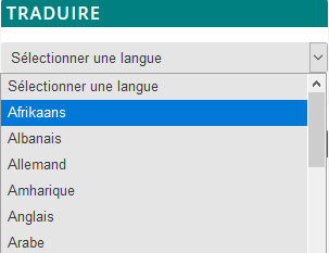
Puis re-selectionner la langue et choisir Français     

Léa Montégut

Roles Conceptualization, Data curation, Investigation, Methodology,
Writing – original draft, Writing – review & editing


Affiliation
Department of Chemical Engineering, Research Laboratory

in Applied Metabolic Engineering, École Polytechnique de Montréal,
Montréal, Québec, Canada

Pablo César Martínez-Basilio

Roles Conceptualization, Methodology


Affiliation
Department of Chemical Engineering, Research Laboratory

in Applied Metabolic Engineering, École Polytechnique de Montréal,
Montréal, Québec, Canada

Jorgelindo da Veiga Moreira

Roles Conceptualization


Affiliation
Department of Chemical Engineering, Research Laboratory
in Applied Metabolic Engineering, École Polytechnique de Montréal,
Montréal, Québec, Canada

Laurent Schwartz

Roles Conceptualization


Affiliation Assistance Publique des Hôpitaux de Paris, Paris, France

Mario Jolicoeur

Roles Conceptualization, Formal analysis, Funding acquisition,
Investigation, Methodology, Project administration, Supervision, Writing – original draft, Writing –
review & editing


mario.jolicoeur@polymtl.ca


Affiliation
Department of Chemical Engineering, Research Laboratory
in Applied Metabolic Engineering, École Polytechnique de Montréal,
Montréal, Québec, Canada