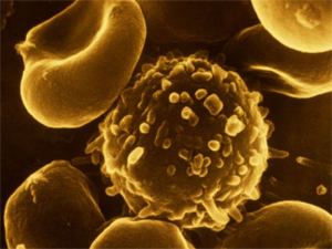
Témoignage – Lymphome de bas grade
Bonjour, Je me permets de vous contacter, d’abord pour vous remercier de vos recherches, mais...
En savoir plus
Bonjour, Je me permets de vous contacter, d’abord pour vous remercier de vos recherches, mais...
En savoir plus
Chers amis, La Fondation Guérir du Cancer vient de publier un point d’activité, et certains...
En savoir plusCe travail (Montégut et al. 2020), fait en collaboration avec le Dr. Laurent Schwartz M.D. et...
En savoir plus
Cet article relate une expérience personnelle, il ne doit en aucun cas être pris comme...
En savoir plus
Je ne peux que remercier grandement le Dr Schwartz. Résumé de l’évolution et des traitements d’un...
En savoir plus
|
Léa Montégut Roles Conceptualization, Data curation, Investigation, Methodology,
|
|
Pablo César Martínez-Basilio Roles Conceptualization, Methodology
|
|
Jorgelindo da Veiga Moreira Roles Conceptualization
|
|
Laurent Schwartz Roles Conceptualization Affiliation Assistance Publique des Hôpitaux de Paris, Paris, France |
|
Mario Jolicoeur Roles Conceptualization, Formal analysis, Funding acquisition,
|